

Management of pelvic chondrosarcoma
1 Carol Davila University, Department of Orthopedics, Bucharest, Romania
2 St. Pantelimon Hospital, Department of Orthopedics, Bucharest, Romania
Abstract
The partial or complete excision of the hemipelvis with sparing of the lower limb is an option of the treatment of pelvic chondrosarcoma and a therapeutic alternative of the interilio-abdominal disarticulation. The operation has in principle the same indications as the interilio-abdominal disarticulation and offers a good solution for avoiding a mutilating operation. The 149 cases include: 120 biopsies, 29 excisional biopsies, 6 interilio-abdominal disarticulations and 14 resections – reconstruction’s, one of with prosthetic reconstruction. The prognostic score was established by assessing: the surgical stage, the site of the tumor, the surgical margins of the tumor, the functional mobility and the postoperative activity. The wide excision of the tumor, a stable reconstruction and an efficient recovery are essential for a successful treatment of pelvic chondrosarcoma. The limb sparing resection-reconstruction represents a highly surgical demanding procedure, followed up by complications in 60% of the cases, so that should be performed only by high skilled surgeons. Hemipelvectomy still remains a well-established life-saving surgery method for patients suffering from vast oncological extensions, where a pelvic resection is not an option.
1. Introduction
Both metastatic and primary tumors of the pelvis usually reach significant mass before diagnostic, due to the large size and compliance of the pelvic cavity. Clinically, most patients do not express any signs or symptoms until late tumor development. Occasionally, a large, asymptomatic mass is felt on abdominal or pelvic examination.
Until late 1970s most pelvic tumors were treated by hemipelvectomy, a procedure often associated with a high percentage of complications and increased rate of mortality [1, 2]. With the advancement in technology, more accurate techniques for imaging of the pelvis, upgraded methods of resections such as internal hemipelvectomy, use of neoadjuvant chemotherapy and radiotherapy, along with improved prosthetic reconstruction, limb-sparing procedures are now performed in the majority of these cases [2-4].
The increased incidence of secondary tumors concomitantly with the lifetime prolongation and with a more reliable oncological detection led to the increase of the number of pelvic tumors generally, under the conditions in which the rate of malignant primitive tumors remains constantly at 15% of the skeletal tumors [4, 5].
The development particularities of the pelvic malignant tumors are related to the structure of the pelvic girdle formed of relatively thin bones from which the extra pelvic of a tumor to the adjacent musculature or its intrapelvic extension (to the viscera of the lesser pelvis) is easily possible. This extension is not limited by septa or important fasciae, neither by other anatomical barriers. The intrapelvic extension manifests itself by a poor clinical symptomatology, explaining the great size of the tumor at the moment of detection [5, 6].
Pelvic chondrosarcoma is not chemotherapy responsive or radiotherapy sensitive, but it is a surgically treatable borderline tumor [4-7].
1.1. Anatomic considerations
A rigorous knowledge of the pelvic and tight anatomy is indispensable in order to apply one of these demanding surgical techniques, the main goal is minimizing both intraoperative and postoperative mortality. A single imaging technique does not offer sufficient data for a tumoral extent to be diagnosed correctly. Data from two or more imaging modalities is required in order to permit a realistic view of the exact anatomic extent [1, 2, 8].
1.2. Musculoskeletal anatomy
The iliac crest, which can be easily palpated, is the attachment site for the abdominal wall musculature, quadratus lumborum and iliacus muscle, the later covering the inner aspect of the ilium.
The acetabulum provides the upper-medial mechanical support of the hip joint. No muscle attachment connects to the acetabulum.
Hip adductors take their origin from the inferior aspect of the pubis. The neurovascular bundle runs along the anterior aspect of the pubis [7, 9].
1.1.1. Neurovascular anatomy
Sacral nerve roots are located inside the vertebral column at the level of L1, where the cauda equina begins, then descending to the sacrum bone. It is considered an unresectable tumor, any type of tumor that penetrates the sacrum beyond the midline, morbidity surpassing the oncologic benefit from the surgery for a procedure at this site [10].
1.1.2. Surgical resection techniques
Pelvic sarcomas are treated either with curettage and cemented hardware reconstruction, by wide resections or by hemipelvectomy. The last two can be classified into two groups: 1) limb sparing resection (pelvic resection) and 2) hemipelvectomy (hindquarter amputations) [11].
Pelvic resection is a term defining a series of techniques grouped together and classified by Enneking. The classification is based on the resected region of the targeted bone: type I, ilium; type II, periacetabular region; type III, pubis; type IV, en bloc resection of the posterior ilium. En bloc resection of the posterior ilium can also be classified as an extended type I resection [12].
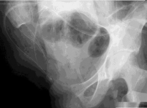
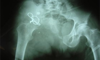
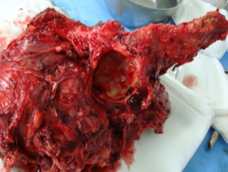
Hemipelvectomy (hindquarter amputations) are required in some extreme cases where a limb-sparing surgery is not a viable option (Figure 1). In Classic Hemipelvectomy (standard) the pubic symphysis and sacroiliac joint are disconnected, common iliac vessels are divided followed by the removal of the pelvic ring and closure with a posterior fasciocutaneous flap. In Modified Hemipelvectomy (conservative) the hypogastric vessels and the inferior gluteal vessels are spared. Extended hemipelvectomy consists in a hemipelvis resection extending the boundaries of the tumor involving the sacroiliac joint and Anterior Flap Hemipelvectomy (described by William Enneking) [13].
Objectives
The experience of our clinic in the treatment of pelvic tumors during a 5-year period is referred in this material. Partial or complete hemiplevic excision is a viable alternative to hemipelvectomy.
2. Materials and Methods
The 149 cases include: 120 biopsies, 29 excisional biopsies, 6 interilio-abdominal disarticulations and 14 resections – reconstructions, one of with prosthetic reconstruction (Figure 1).
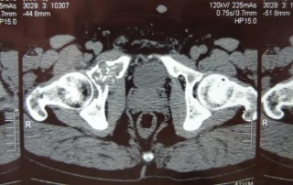
Angio- magnetic resonance (Angio- MRI), scintigraphy, computed tomography (CT) and radiographic investigation in classical incidence and oblique incidence where used as methods for the preoperative tumoral staging (Figure 3).
The preoperative status was established by means of a multidisciplinary investigation consisting in:
Assessment of the clinical state: anamnesis, tumoral development rate and drug addiction.
Biological investigation: hematological analysis and serum biochemistry, tumoral markers, alkaline phosphatase, phosphocalcium balance, anesthetic and surgical risk factors.
Study of the tumor type: 2 cases of chondrosarcomas (1B), 1 malignant fibroma histiocytoma, 1 pelvic hydatid cyst.
Other tumoral locations investigated by scintigraphy and pulmonary CT.
Study of the response to the treatment
Two criteria where used for the inclusion of the patients in the category of resection-reconstruction treatment:
Obtaining adequate surgical margins using this procedure would be possible.
The margin obtained by interilio-abdominal amputation would not have been oncological safer than that obtained by resection-construction.
The site of the tumor was in two cases in the II +1 region, in one case in the region II and in one case in the region I +II +III.
The preoperative preparation protocol included: two-days-preparation of the digestive tract by liquid diet and antibiotic therapy including Cefamandole, Gentamicin and Metronidazole for the intestinal flora sterilization. A urethral catheter was used for marking the ureter in order to avoid its intraoperative lesion. General and through epidural catheter anesthesia was used and the monitoring consisted in invasive arterial tension, SAO2, ETCO2, ECG, urinary output per hour, control venous line and central temperature.
1.3. Surgical techniques
A standard ilio-inguino-perineal incision was used as the main resection method with anterior, posterior or lateral extensions in conformity to the tumoral site, followed by the iliac, gluteal and obturator vessels control, assisted by the vascular surgery team. Pelvic cavity inspection was possible after the disinsertion of the abdominal musculature from the iliac and the pubic bone was completed. Posterior, the region of the sciatic notch and the sciatic nerve were laid bare, the resection lines could be identified and 3 osteotomies according to the planning were carried out, including the hip joint when the tumor has invaded the articular cavity. In one case the surgical margins obtained were non-contaminated (chondrosarcoma), in two cases the margins were contaminated (malignant fibromas, histiocytoma and chondrosarcoma), whereas in another case, strict oncological problems were not involved [14].
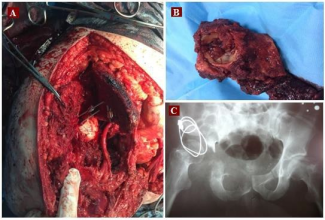
The reconstruction technique included three ilio- femoral pseudo-arthrosis and one reconstruction through the Paget technique with total hip joint. The ilio-femoral pseudo-arthrosis reconstruction was achieved by anchorage of the proximal end of the femur to the iliac wing, according to the type II/III or IIA/III resections with intra-acetabular invasion (Figure 4, 5, 6).
3. Results
Pelvic tumors surgery was associated with a great number of complications, as 3 out of 4 patients who underwent resection-reconstruction were recorded with one or several complications post operatory.
The most common complication, the infection with or without dehiscence, was present in 2 patients. In the case of the patient with hydatid cyst, the staphylococcus infection was treated by early debridement and antibiotic therapy.
In one of the 2 cases in which the surgical margins were considered contaminated, more precisely, the patient with the malignant fibrous histiocytoma, a recent scrotal edema occurred on the operated side suggesting a local recurrence.
One out of 4 patients, in whom the Paget resection-reconstruction technique was applied, presented neurological complication. The paralysis of the peroneal nerve might have occurred while total hip prosthesis was applied, to which the femoral part was fitted with a long neck, tensioning the sciatic nerve.
Other complications were present: in two patients a moderate lumbalgia was present; one patient manifested a thrombophlebitis and another patient, a cutaneous necrosis. No visceral lesions were detected in any of the patients.
1.4. Evaluation of the reconstruction
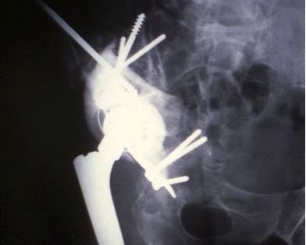
Post operatory pelvic instability was present in all the studied cases, loss of the articular connection between the femur and the sacroiliac joint on the resected side. The Puget arthroplasty reconstruction technique required a 9 cm resection from the proximal extremity of the femur, which was used as a graft, restoring the anatomical pelvic continuity between the superior pelvic ramus and the sacroiliac joint, followed by the acetabular prosthetic component implantation. The proximal femur was reconstructed by using a Kent type prosthesis (Figure 7, 8). Post operatory, six weeks immobilisation was achieved with the use of a pelvipodal plaster cast.
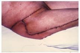
In conformity to the Musculoskeletal Tumor Society, pain, mobility, stability, deformation, muscle strength, physiological tolerance and functional activity were analyzed in the context of the patient’s functional assessment evaluation. For each parameter 5 points were assigned at the most, 35 points corresponding to a 100% value. The average score of the 4 cases was of 21 points, representing a value of 60% which corresponds to a satisfactory score.
4. Discussion
Although the surgery of pelvic bone tumors achieved exceptional advances due to the advancement in the imaging investigation sector, prosthetic reconstruction, anesthesia and upgraded methods of resection, it is still a technically demanding procedure, followed up by numerous complications. Most complications are the result of the tumor abscission, compared with the lower rate of complication resulting from reconstruction. Literature data confirms that this type of intervention is burdened with 60% complications [15].
In some extreme cases, such as vast tumoral extensions, hemipelvectomy still remains a lifesaving procedure.
During the resection-reconstruction by iliofemoral pseudarthrosis 11 blood units were transfused and 19 blood units for the Paget type resection-reconstruction with total hip prosthesis. The duration of the operation was 6.5 for the iliofemoral pseudarthrosis reconstruction respectively 11 hours for the Paget procedure [16].
The remaining mass of the abductor muscle represents a decisive element in the mobility function post resection.
Modern day techniques aim at either a biological or mechanical reconstruction [17].
For the mechanical reconstruction, an individualized pelvic implant is used along with a total hip prosthesis. Although the promoters of this method consider that it assures the best functional results, this reconstruction procedure comes with a series of specific complications such as: transosseous iliac migration and dislocation [18].
Frozen osteochondral allografts are used in the biological reconstruction procedure, retaining the length of the limb and the articular mobility. Nonetheless, this surgical method comes with frequent complications: interface consolidation, infection and fatigue fracture [19].
5. Conclusions
Limb-sparing pelvic resection-reconstruction consists in a remarkable result on patients whom a hemipelvectomy procedure would not offer better oncological results. Limb sparing resection- reconstruction represents a highly surgical demanding procedure, followed up by complications in 60% of the cases, so that should be performed only by high skilled surgeons.
Hemipelvectomy still remains a well-established life-saving surgery method for patients suffering from vast oncological extensions, where a pelvic resection is not an option. Multidisciplinary staging, imaging, biochemistry, clinical and, last but not the least, surgical fields are required in order to obtain a high quality result.
References
- Abudu A, Grimer RJ, Cannon SR, Carter SR, Sneath RS. Reconstruction of the hemipelvis after the excision of malignant tumours. Complications and functional outcome of prostheses. J Bone Joint Surg Br. 1997; 79(5): 773-9. PMID: 9331034 https://doi.org/10.1302/0301-620X.79B5.6749
- Arvinius C, González-Pérez A, García-Coiradas J, García-Maroto R, Cebrián-Parra JL. Paraneoplastic vasculitis associated to pelvic chondrosarcoma: a case report. SICOT J. 2016; 2: 8. PMID: 27163097 https://doi.org/10.1051/sicotj/2015042
- Delepine F, Delepine G, Sokolov T, Hernigou P, Goutallier D. Resultats des prostheses composites au cement apres resection acetabulaire pour sarcome. Rev Chir Orthop Reparatrice Appar Mot. 2000; 86(3): 265-77. PMID: 10844357
- Ozturker C, Incedayi M, Kara K, Sonmez G. Giant pelvic chondrosarcoma with vertebral column invasion. Spine J. 2016; 16(4): e239-40. PMID: 26515393 https://doi.org/10.1016/j.spinee.2015.10.031
- Enneking WF, Spanier SS, Malawer MM. The effect of the anatomic setting on the results of surgical procedures for soft parts sarcoma of the thigh. Cancer 1981; 47(5): 1005–22. PMID: 7226034 https://doi.org/10.1002/1097- 0142(19810301)47:5<1005::AID-CNCR2820470532>3.0.CO;2-9
- Outani H, Hamada K, Imura Y, Oshima K, Sotobori T, Demizu Y, Kakunaga S, Joyama S, Imai R, Okimoto T, Naka N, Kudawara I, Ueda T, Araki N, Kamada T, Yoshikawa H. Comparison of clinical and functional outcome between surgical treatment and carbon ion radiotherapy for pelvic chondrosarcoma. Int J Clin Oncol. 2016; 21(1): 186-93. PMID: 26150259 https://doi.org/10.1007/s10147-015-0870-z
- Arnal-Burró J, Calvo-Haro JA, Igualada-Blazquez C, Gil-Martínez P, Cuervo-Dehesa M, Vaquero- Martín J. Hemipelvectomy for the treatment of high-grade sarcomas: Pronostic of chondrosarcomas compared to other histological types. Rev Esp Cir Ortop Traumatol. 2016; 60(1): 67-74. PMID: 26091572 https://doi.org/10.1016/j.recot.2015.04.002
- Gillis CC, Street JT, Boyd MC, Fisher CG. Pelvic reconstruction after subtotal sacrectomy for sacral chondrosarcoma using cadaveric and vascularized fibula autograft: Technical note. J Neurosurg Spine. 2014; 21(4): 623-7. PMID: 25084027 https://doi.org/10.3171/2014.6.SPINE13657
- Possover M, Uehlinger K, Ulrich Exner G. Laparoscopic assisted resection of a ilio-sacral chondrosarcoma: A single case report. Int J Surg Case Rep. 2014; 5(7): 381-4. PMID: 24862027 https://doi.org/10.1016/j.ijscr.2014.04.007
- Blakeney WG, Day R, Cusick L, Smith RL. Custom osteotomy guides for resection of a pelvic chondrosarcoma. Acta Orthop. 2014; 85(4): 438- 41. PMID: 24847792 https://doi.org/10.3109/17453674.2014.920988
- Espejo-Sánchez G, Rico-Martínez G, Linares- González LM, Delgado-Cedillo E, Clara- Altamirano MA. Periacetabular pelvic reconstruction for chondrosarcoma with autograft of the proximal femur. Acta Ortop Mex. 2012; 26(4): 250-4. PMID: 23320328
- Mankin HJ, Doppelt SH, Sullivan TR, Tomford WW. Osteoarticular and intercalary allograft transplantation in the management of malignant tumors of bone. Cancer. 1982; 50(4): 613-30. PMID: 7046906 https://doi.org/10.1002/1097- 0142(19820815)50:4<613::AID-CNCR2820500402>3.0.CO;2-L
- Nieder E, Elson RA, Engelbrecht E, Kasselt MR, Keller A, Steinbrink K. The saddle prosthesis for salvage of the destroyed acetabulum. J Bone Joint Surg Br. 1990; 72(6): 1014-22. PMID: 2246283
- Campanacci D, Chacon S, Mondanelli N, Beltrami G, Scoccianti G, Caff G, Frenos F, Capanna R. Pelvic massive allograft reconstruction after bone tumour resection. Int Orthop. 2012; 36(12): 2529- 36. PMID: 23090357 https://doi.org/10.1007/s00264-012-1677-4
- Gaston CL, Sumathi VP, Grimer RJ. Is it ever safe to discharge a chondrosarcoma of pelvis? Report of a local recurrence after 10 years. Musculoskelet Surg. 2014; 98(3): 241-6. PMID: 22990984 https://doi.org/10.1007/s12306-012-0224-1
- Puget J, Utheza G. Reconstruction de l'os iliaque a l'aide du femur homolateral apres resection d'une tumeur pelvienne. Rev Chir Orthop Reparatrice Appar Mot. 1986; 72(2): 151-5. PMID: 3715099
- Uchida A, Myoui A, Araki N, Yoshikawa H, Ueda T, Aoki Y. Prosthetic reconstruction for periacetabular malignant tumors. Clin Orthop Relat Res. 1996; (326): 238-45. PMID: 8620647 https://doi.org/10.1097/00003086-199605000- 00029
- Angelini A, Guerra G, Mavrogenis AF, Pala E, Picci P, Ruggieri P. Clinical outcome of central conventional chondrosarcoma. J Surg Oncol. 2012; 106(8): 929-37. PMID: 22649023 https://doi.org/10.1002/jso.23173
- Mavrogenis AF, Gambarotti M, Angelini A, Palmerini E, Staals EL, Ruggieri P, Papagelopoulos PJ. Chondrosarcomas revisited. Orthopedics. 2012; 35(3): e379-90. PMID: 22385450 https://doi.org/10.3928/01477447- 20120222-30
