

Parasagittal meningiomas – literature review and a case report
1 Carol Davila University, Department of Neurosurgery, Bucharest, Romania
2 St. Pantelimon Emergency Clinical Hospital, Department of Neurosurgery, Bucharest, Romania
3 Ploiești County Emergency Hospital, Department of Neurosurgery, Ploiești, Romania
Abstract
Meningiomas are tumors that can develop anywhere along the neuraxis, but with increased concentration in some specific areas. Parasagittal meningiomas have the dural attachment on the external layer of the superior sagittal sinus (SSS) and invade the parasagittal angle displacing brain away from its normal position.
Among meningiomas, the parasagittal location is the most common (22%). Taking into account their anatomic insertion along SSS, parasagittal meningiomas can have their dural attachment in the anterior, the middle or the posterior third of the SSS. Most frequently parasagittal meningiomas are located in the middle third of the superior sagittal sinus (between coronal suture and lambdoid suture). The clinical picture of parasagittal meningiomas depends on the tumor location along the SSS and so is the attitude towards ligation and reconstruction of the sinus. Controversial issues regarding surgical management of parasagittal meningiomas concerning leaving a tumor remnant that invades the SSS instead attempting total resection, or the attitude in the case of totally occluded segment of a sinus are summarized in this paper. The special care for the venous system is emphasized. The recurrence matter is also approached underlining the importance of adjuvant radiosurgery for the management of residual tumors. Results described in the main papers of the literature are reviewed.
Conclusions are referring to the historical evolution regarding the surgical management of parasagittal meningiomas: aggressiveness of resection, sinus reconstruction, importance of adjuvant techniques: radiosurgery, endovascular surgery and to the importance of microsurgery and careful and meticulous planning of the approach in order to avoid interference with venous collaterals. A suggestive clinical case from the authors experience is presented.
1. Introduction
Meningiomas are tumors with the origin in the arachnoid cap cells. This type of meningeal cells can be found anywhere along the neuroaxis, but with an increased concentration in some specific areas. As a consequence, a meningioma can develop anywhere in the central nervous system but there is a higher incidence in elective locations. Any meningioma has a dural attachment and based on this feature we can classify meningiomas in distinct groups.
2. Discussion
1.1. Anatomy
The sagittal sinus has a triangular shape in coronal section and his size gradually increases as it extends posterior [1]. The superior sagittal sinus communicates with irregular venous cavities called lateral venous lacunae, which lie in the dura mater. In certain areas, the arachnoid projects into the venous sinuses to form the arachnoid villi. These are mushroom-shaped outpouchings of the subarachnoid space that invaginate into them allowing CSF to flow inside the sinuses. When they are very large, the villi can be perceived with naked eye and are called Pacchioni’s granulations. Sometimes these granulations may be seen on magnetic resonance imaging as a filling defect or a mass within a large dural sinus [2].
Parasagittal meningiomas have the dural attachment on the external layer of the SSS and invade the parasagittal angle displacing brain away from its normal position. Cushing and Eisenhardt, in their series of 295 meningiomas, found that the parasagittal location is the most common, 22% [3]. The frequency of parasagittal meningiomas among all meningiomas ranges in the literature between 16.8 and 25.6% [4]. Olivecrona classified parasagittal meningiomas taking into account their anatomic insertion along SSS. He divided the superior sagittal sinus into anterior, middle and posterior third having in mind the different consequences of sinus occlusion in each area [5]. Most frequently parasagittal meningiomas have the dural attachment in the middle third of the superior sagittal sinus (between coronal suture and lambdoid suture). This location is found in 37 - 70% of all parasagittal meningiomas. Along the first third of the SSS (from crista galli to the coronal suture) are located 15 - 42% of parasagittal meningiomas and only 9% to 16% of parasagittal tumors are located along the posterior third of the SSS (between the bregma and torcula) [6].
2.1. Signs and symptoms
The clinical picture of parasagittal meningiomas depends on the tumor location along the SSS. Thus, all tumors can produce headache, but meningiomas of the anterior third of the SSS often cause personality changes, tumors of the middle third of the SSS are often associated with Jacksonian seizures and progressive hemiparesis and those of the posterior third can cause hemianopsia [3-5].
It is generally accepted that, whilst the anterior third can usually be resected without morbidity, ligating the middle or posterior third of the sinus carries significant risk of venous infarction [6, 7].
There are also some other anatomic considerations that can be made regarding topographical differences among parasagittal meningiomas. Thus, the sagittal sinus is narrower in its anterior half and the veins entering the sinus are fewer in number and smaller than those behind the coronal suture, making the surgical approach easier in the anterior half. Moreover, the growth of tumors of the middle third of the parasagittal sinus against the paracentral lobule and the precentral/ postcentral gyri around the central sulcus makes them clinically evident early, explaining why they do not have the time to expand in size as do the tumors in the other two thirds [8, 9].
1.2. Surgical management
Surgery of meningiomas involving the sagittal sinus challenges the surgeon with a dilemma: to leave the invasive fragment having a higher rate of recurrence or to attempt total removal putting at risk the venous circulation [9, 10]. Current tendency nowadays is resection of the meningioma outside the walls of the sinus with coagulation of the tumor remnant, followed if needed by an “en bloc” removal of the residual tumor when complete sinus occlusion occurs [10, 11].
All neurosurgeons operating parasagittal meningiomas emphasize the importance of preserving the bridging veins [1, 8, 10], especially the ones of the group draining in the middle third of the sagittal sinus.
Another matter of almost general consensus is that surgeons have to avoid interrupt a sinus that is partially occluded. On the other hand, sometimes, a complete resection of a totally occluded segment of a sinus can lead to brain swelling and venous infarction if venous collateral circulation is affected. For this reason, a surgical intervention for a parasagittal meningioma that totally occludes the SSS must be carefully planned and performed, taking into account that even superficial or intra-diploic collaterals can be of paramount importance for the cerebral venous drainage [12, 13].
Even in extensive resections with sinus reconstruction or bypass that raised perioperative morbidity and mortality, local recurrence still remains a major problem [8, 9, 11]. Therefore, because of the potential morbidity of such cases and the efficacy of adjuvant radiosurgery for the management of residual tumors and recurrent meningiomas, surgeons have adopted lately a more conservative approach in the management of sinus invasion [14].
2.2. Outcome and results
In Olivecrona’s series, of 196 parasagittal meningiomas resected without sinus reconstruction, morbidity was 12.3%, half of which being attributed to venous damages [5]. The mortality rate was 10%, with significant differences depending on location: 12.8% for the middle third, 9% for the posterior third and 5.3% for the anterior third of the sinus. In Bonnal and Brotchi’s series of 21 cases, mortality was 4.7%, namely 1 patient [13]. In another recent report of 108 cases, brain swelling was found in nine patients (8.9%).
The postoperative mortality in cases with total tumor removal has obviously improved since the first attempts of complete resections performed several decades ago. Improvement in microsurgical techniques together with sinus repair when total tumor removal is performed has significantly changed the outcome of these patients. In Sindou series of 100 cases, mortality was 3%, three patients dying from postoperative brain swelling [11, 15]. These patients had extensive resection of the totally invaded segment of the sinus without venous flow restoration.
In the same series there were 15 sinus repairs with dura or fascia from which 13 were confirmed angiographically to be patent. In the authors experience, the preferred material was the thin and glossy fascia temporalis. When a synthetic graft was used, none of them remained patent in spite of anticoagulation therapy. In consequence, authors don’t recommend the use of synthetic grafts for repair of the venous system. If temporary occlusion of the sinus is required, the use of pledgets of Surgicel in the sinus lumen and at the ostia of the afferent veins is suggested. This technique is considered to be better that the use of aneurysm clips or balloons which were even found to damage the sinus walls. Silicone tubes used as shunts don’t pass well through the sinus lumen because of the presence of septae, especially in the middle third of the SSS [14-16].
1.3. Case report
A 43-year old woman presented with a long history of headache (approximately 5 months) and a recently installed mild right-side hemiparesis. The neurological examination did not reveal any other focal deficits besides the right hemiparesis, which was predominantly brachial. Babinski’s sign was positive on the right side.
Enhanced CT imaging revealed a left frontal extra-axial, 55 x 51 x 47mm in size, isodense, contrast-enhanced well-defined mass, adjacent to the falx cerebri and surrounded by cerebral edema. The lesion was non-homogeneous, presenting small hypodense areas inside. It had a mass effect on the ventricular system, shifting the midline by approximately 15mm to the right (Figures 1, 2).

Operation: a left frontal-parietal craniotomy was made, slightly crossing the midline to the right. A semicircular dural incision was made, leaving a median pedicle, revealing an extra-axial tumor adhering to the superior sagittal sinus and falx cerebri, with variable consistency (Figure 3).
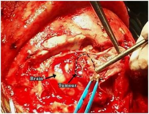
The lesion was well vascularized and it had a spherical shape, with a diameter of approximately 5-6 cm. The tumor was then removed in gross-total fashion, by intracapsular debulking. The tumor capsule was quasi-totally resected, leaving in place an approximately 1cm by 1cm residue which was adherent to the superior sagittal sinus. A Simpson IV grade resection was noted [17, 18].
Postoperatively, a non-enhanced CT-scan, performed one week after the surgical intervention showed gross-total resection with a left parasagittal hypodensity, slightly non-homogeneous, with small haematic areas and minimal pneumocephalus (Figures 4, 5).

A minor improvement in the right hemiparesis was observed, with no other neurological deficits installed after surgery. The patient was discharged 7 days postoperatively, with an almost normal neurological status [19].
3. Conclusions
The treatment of parasagittal meningiomas has evolved during time from aggressive gross total removal with flow restoration using bypass techniques and sinus reconstruction to multimodal approaches involving microsurgery, endovascular surgery, and radiosurgery. This evolution has significantly decreased surgical mortality and morbidity and has improved outcomes, but the long-term effects of the new above-mentioned therapies are still to be assessed [13, 14].
The goals of surgical treatment are to achieve a tumor resection as complete as possible without producing neurological impairment or affecting the quality of life. Although, at this moment, several venous flow restoration techniques are available to surgeons, their efficacy and safety have not been validated yet in multicentric randomized studies, and they seemingly expose patients to added morbidity, especially if performed in inexperienced centers.
Although the resection of completely occluded sinuses can be considered a safe strategy, studying the collateral venous flow must not be neglected. Careful and meticulous planning of the approach in order to avoid interference with venous collaterals is mandatory [15]. Stereotactic radiosurgery is an useful tool, allowing for treatment of residual tumors or recurrent disease. Other adjuvant technologies will also contribute more and more in the future to the management of these challenging lesions [16].
References
- Sindou M, Auque J. The intracranial venous system as a neurosurgeon’s perspective. Adv Tech Stand Neurosurg. 2000; 26:131–216. PMID: 10997200 https://doi.org/10.1007/978-3-7091- 6323-8_5
- Alghamdi M, Li H, Olivotto I, Easaw J, Kelly J, Nordal R, Lim G. Atypical Meningioma: Referral Patterns, Treatment and Adherence to Guidelines. Can J Neurol Sci. 2017; 44(3): 283-287. PMID: 28488953 https://doi.org/10.1017/cjn.2016.449
- Zakhari N, Torres C, Castillo M, Nguyen TB. Uncommon Cranial Meningioma: Key Imaging Features on Conventional and Advanced Imaging. Clin Neuroradiol. 2017; 27(2): 135-144. PMID: 28466126 https://doi.org/10.1007/s00062-017- 0583-y
- Fernandez C, Nicholas MK, Engelhard HH, Slavin KV, Koshy M. An analysis of prognostic factors associated with recurrence in the treatment of atypical meningiomas. Adv Radiat Oncol. 2016; 1(2): 89-93. PMID: 28740874 https://doi.org/10.1016/j.adro.2016.03.001
- Hoessly GF, Olivecrona H. Report on 280 cases of verified parasagittal meningioma. J Neurosurg. 1955; 12(6): 614–25. PMID: 13272053 https://doi.org/10.3171/jns.1955.12.6.0614
- Kang JK, Jun SS, Sung WH, et al. Surgical management of meningioma involving the superior sagittal sinus. Surgery of the intracranial venous system. In: Hakuba A, ed. New York: Springer, 1996:252–259.
- Hilmani S, Houass Y, El Azhari A. Paraventricular meningioma revealed by mental disorder. Surg Neurol Int. 2016; 7(Suppl 40): S1004-S1007. PMID: 28144473 https://doi.org/10.4103/2152- 7806.195580
- Sindou M, Auque J. The intracranial venous system as a neurosurgeon’s perspective. Adv Tech Stand Neurosurg. 2000; 26: 131–216. PMID: 10997200 https://doi.org/10.1007/978-3-7091- 6323-8_5
- Ahmed AK, Morrison JF, Lakis NS, Stopa E, Doberstein C. Atypical meningioma mimicking high-grade glioma. Neurology. 2016; 87(21): 2281-2. PMID: 27872223 https://doi.org/10.1212/WNL.0000000000003364
- Shim Y, Lee SH. Failed First Craniotomy and Tumor Removal of Parasagittal Meningioma with Severe Peritumoral Brain Edema. Brain Tumor Res Treat. 2016; 4(2): 124-127. PMID: 27867923 https://doi.org/10.14791/btrt.2016.4.2.124
- Sindou MP, Alvernia JE. Results of attempted radical tumor removal and venous repair in 100 consecutive meningiomas involving the major dural sinuses. J Neurosurg. 2006; 105(4): 514–25. PMID: 17044551 https://doi.org/10.3171/jns.2006.105.4.514
- Lim SH, Moon SH, Kim MM. Incidental frontoparietal parasagittal meningioma in a patient with acute bilateral optic neuritis. Int J Ophthalmol. 2014; 7(6): 1064-6. PMID: 25540766
- Hambra DV, Danilo de P, Alessandro R, Sara M, Juan GR. Meningioma associated with acute subdural hematoma: A review of the literature. Surg Neurol Int. 2014; 5(Suppl 12): S469-71. PMID: 25422791 https://doi.org/10.4103/2152- 7806.143724
- Sughrue ME, Sayegh ET, Parsa AT. Towards a hypermodern theory of meningioma surgery. Clin Neurol Neurosurg. 2014; 126: 69-75. PMID: 25215444 https://doi.org/10.1016/j.clineuro.2014.06.002
- Oka K, Go Y, Kimura H, Tomonaga M. Obstruction of the superior sagittal sinus caused by parasagittal meningiomas: the role of collateral venous pathways. J Neurosurg. 1994; 81(4): 520– 4. PMID: 7931584 https://doi.org/10.3171/jns.1994.81.4.0520
- Brain meningioma with initial manifestation similar to cervical radiculopathy: a case report. Huang YH, Hong CZ, Wu WT, Li KT, Chou LW. Neuropsychiatr Dis Treat. 2014 Jun 25;10:1175- 81. PMID: 25028552
- Voultsinou D, Matis GK, Chrysou OI, Birbilis TA, Cheva A, Geroukis T. An atypical meningioma demystified and advanced magnetic resonance imaging techniques. J Cancer Res Ther. 2014; 10(2): 387-9. PMID: 25022401 https://doi.org/10.4103/0973-1482.136666
- Nowak A, Dziedzic T, Czernicki T, Kunert P, Marchel A. Surgical treatment of parasagittal and falcine meningiomas invading the superior sagittal sinus. Neurol Neurochir Pol. 2014; 48(3): 174-80. PMID: 24981181 https://doi.org/10.1016/j.pjnns.2014.05.003
- Andric M, Dixit S, Dubey A, Jessup P, Hunn A. Atypical meningiomas--a case series. Clin Neurol Neurosurg. 2012; 114(6): 699-702. PMID: 22285882 https://doi.org/10.1016/j.clineuro.2011.11.023
