

Comparative evaluation of primary repair method with Z-plasty and Limberg flap technique on pilonidal sinus treatment of children
1 SBU KOCAELI DERINCE TRAINING AND RESEARCH HOSPITAL, DEPARTMENT OF PEDIATRIC SURGERY, DERINCE, KOCAELİ, TURKEY
Abstract
Introduction. Although numerous surgical closure methods have been proposed for the treatment of Pilonidal Sinus Disease (PSD), the ideal treatment method has not yet been identified due to high recurrence rates. The objective of this study is to compare the closure technique that uses traditional post-pilonidal sinus excision Primary Repair (PR) method with closure technique that uses flap-based surgical methods such as Z-plasty (ZP) and Limberg flap (LF) in terms of their role in ensuring healing and their complication and recurrence rates. Materials and Methods. 61 patients (39 male and 22 female) who underwent surgery for PSD using the traditional Primary Repair, flap-based Limberg flap, and Z-plasty flap techniques were included in the study. The median age of the patients was 16.2 years, and the follow-up period was between 3 to 6 months. Results. Throughout the follow-up period, there developed recurrence in two patients and wound dehiscence and wound site infection in 6 patients in the PR group, whereas recurrence was observed in one patient and wound dehiscence in two patients in the LF group. While one patient developed hematoma, and one patient developed seroma in the ZP group, no recurrence or flap necrosis was observed in the ZP group. Discussions. After comparing complication frequency, recurrence rate, and cosmetic evaluation results, it was understood that the results belonging to LF and ZP groups, which represent flap-based surgical methods, were statistically more significant than the PR method group (p<0.05). When comparing flap-based surgical closure methods among themselves, it was seen that the surgical recurrence, complication and cosmetic results of ZP and LF closure methods were statistically more significant in favor of the ZP group (p<0.05). Conclusions. Although it requires technical expertise, it is recommended that sinus excision and flap-based surgical methods and the ZP surgical closure technique be used in surgical PSD treatment in pediatric age groups due to low complication and recurrence rates and better cosmetic results.
Introduction
Pilonidal sinus disease is a common disease that is very difficult to manage [1]. According to Monro and McDermott [2, 3], the factors responsible for the development of pilonidal sinus disease (PSD) are believed to be deep birth clefts. Many hair follicles and hair points around these clefts are directed towards the depth of the congenital sacral depression. It is thought that excessive recurrent trauma applied to the sacrococcygeal region causes the formation of pilonidal cysts [4].
Although numerous surgical closure methods have been proposed for the treatment of Pilonidal Sinus Disease (PSD), the ideal treatment method has not yet been identified due to high recurrence rates.
This study aims to compare the surgical complication, recurrence and cosmetic results of the traditional primary repair method (PR) which is used in our clinic for surgical treatment of PSD cases with the Limberg Flap (LF) and Z-plasty flap (ZP) techniques which are flap-based surgical closure methods for treatment.
Materials and Methods
An approval no. 2019-91 dated 26.09.2019 was obtained from the SBÜ Kocaeli Derince Training and Research Hospital Ethics Committee for this retrospective study. The patients who underwent surgical closure treatment for PSD between June 2016 and July 2018 were determined as the study group. All patient BMI values were calculated, and routine laboratory studies were carried out prior to surgical operation. Patients' sinus openings in the sacral region and their distance from the anal region were evaluated preoperatively to determine which type of flap-based surgery would be applicable. All patients were evaluated to be in the ASA 1 group in terms of anesthesia risk. The related regions were washed with antibacterial soap and depilated the night before the operation. The patients were transferred to the operating room in the morning of surgery. All patients received spinal block anesthesia and placed in the prone position. The area of surgery was prepared and soaked with betadine. After determining the surgical method applicable to the patient, the elliptical incision area for PR and ZP groups and the rhomboid excision area for the LF group and making sure that the areas marked included all sinus openings, the pilonidal sinus region to be excised was established, and the flap lines were determined for Limberg flap or Z-plasty flap (Figure 1).
The pilonidal sinus region was excised through the presacral fascia, and deep granulation tissues were cleared (Figure 2).
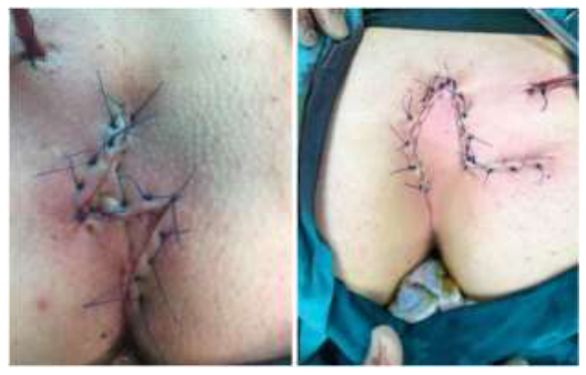
After this procedure, tissues of patients receiving traditional primary closure were primarily interconnected in the midline. The subcutaneous layer was closed off with 2/0 vycril suture and the dermis with 3/0 prolene suture material. For patients receiving treatment with Limberg flap and Z-plasty flap, LF and ZP flaps formed with 45-60-degree angles were prepared following hemorrhage control (Figure 3).
After total excision was completed, the flaps that were formed were interconnected without tension after placement of 1 hemovac drain in the lodge. The subcutaneous layer was sealed off with 2/0 vycril suture and the dermis with a 3/0 prolene suture material.
Patients to whom PR was applied were discharged one day after the operation and the patients who received LF and ZP treatment were discharged one day later after the drainage dropped below 10 cc/day and the drains were removed. All patients were called in for outpatient clinic checkup 2 and 4 weeks after discharge. In the outpatient clinic checkups, another outpatient clinic checkup to be performed 3 months after the operation was recommended to patients without any problem in order to perform a functional and cosmetic evaluation on the operation. Post-op patient satisfaction was categorized as Poor (1 point), Fair (2 points), Good (3 points) and Excellent (4 points). Patients who failed to come in for a checkup were communicated by phone, and their operation-related satisfaction levels and complaints were inquired. Age, gender, operative findings, post-operative complications, incision line complaints in the gluteal region, recurrence status, and operation satisfaction of patients were logged, and the results were subjected to a statistical evaluation using the SPSS 17 software. The values were provided as average and standard deviation.


Results
Sixty-one patients who received surgical treatment for post-pilonidal sinus excision closure using traditional PR, LF, and ZP methods were included in the study. After pilonidal sinus excision, the PR method was used in 16 patients (26.2%), LF method was used in 21 patients (34.4%), and ZP method was used in 24 patients (39.3%) for closure procedure.
The age average of patients included in the study was 16.2 years in the PR group (13.5-17.4), 15.6 years in the ZP group (14.1-17.3) and 16.6 years in the LF group (13.2- 17.5). The body-mass index (BMI) of patients was 25,8 ± 2,1 kg/m2 in the patient group subjected to PR, 26.6 ± 3.76 kg/m2 (21- 36.5) in the patient group subjected to ZP and 25,3 ± 1,8 kg/m2 in the patient group subjected to LF. The duration of surgery was 36,1 ± 11,0 min. (25-48) for the patient group subjected to PR, 73,2 ± 8,3 min. (65-85) for the patient group subjected to the Limberg flap and 53.0 ± 14.7 min. (38-68 min.) for the patient group subjected to ZP. The length of hospital stay was 1.3 ± 0.3 days [1-2], 2.1 ± 0.3 days (1.7-2.4) and 2.6 ± 1.3 [2-4] days for PR, LF, and ZP patient groups, respectively.
Type of Surgery PR 1 ZP 2 LF 3 p Average Age (Year) 16,5 16 16,5 0,88 Sex(F/M) 5/11 9/15 8/13 0,24 BMI 4 (kg/m2) 25,8 ± 2,1 26.6 ± 3.76 25,3 ± 1,8 0,22 Surgery Duration (min.) 36,1 ± 11,0 53.0 ± 4.7 73,2 ± 8,3 0,03 5 Length of hospital stay (day) 1,3 ± 0,3 2,6 ± 1,3 2,1 ± 0,3 0,02 5 Incision Length 5,6 ± 1,4 5,9 ± 1,2 6,1 ± 1,1 0,03 5 Post-op midline incision length 5,8 ± 1,3 9,1 ± 1,1 6,3 ± 1,2 0,01 6 Excision area 21,4 cm2 23,2 cm2 42,4 cm2 0,02 6 Suture removal period (day) 10 12 12 Follow-up period (month) 3-6 3-6 3-6 Complication 6 (%37) 2 (%8,3) 3(%14,2) Recurrence 2 (%12,5) 0 1 (%4,7) 1-PR: Primary Repair; 2-ZP :Z Plasty; 3-LF:Limberg Flap; 4- BMI:Body Mass Index; 5-Among PR and Flap-based surgeries; 6-Among ZP and LF surgeries Wound site infection in three patients, suture dehiscence in three patients and recurrence in two patients in the PR group were identified in the early stages of post-operative follow-up period. Such patients in the PR group who developed wound site infection and suture dehiscence were treated with drainage, wound dressing and antibiotherapy. The wound site of patients whose suture dehiscence continued despite treatment was revised by using local anesthesia. Patients developing recurrence after primary repair were successfully treated by re-operating using the Z-plasty flap method. While 8 patients (50 %) gave 5 points in the cosmetic evaluation performed in the 3rd month after surgery, 4 patients (25 %) gave 4 points, 2 patients (12.5%) gave 2 points and 2 patients (12.5 %) gave 2 points. Four patients complained about pain in the wound site, one patient complained about numbness and five patients complained about stiffness in the 3rd-month post-op checkup. The post-op patient satisfaction in the PR patient group was valued at 2.73 ± 0.88 points. Four patients from the PR patient group complained about soreness in the surgery area one complained about numbness and five complained about stiffness in the post-op satisfaction evaluation.
Three patients in the group to whom Limberg flap method was applied developed wound site infection and two patients developed suture dehiscence in the early post-operative stage. The infection regressed following checkups performed by removing sutures and dressing wounds on a regular basis. Two patients developing suture dehiscence received wound revision by local anesthesia after regular wound dressing and antibiotherapy. One patient developed recurrence and was successfully treated by undergoing a re-operation using the Z-plasty flap procedure. While 13 (61.9 %) patients from the LF group gave 4 points in the post-op checkups; 4 patients (19.0 %) gave 3 points, 3 patients (14.2) gave 2 points and the 1 patient developing a recurrence (4.7 %) gave 2 points. The post-op patient satisfaction in the LF group was valued at 3.06 ± 0.70 points. Two patients from the LF patient group complained about soreness in the surgery area, four patients complained about numbness and three patients complained about stiffness in the post-op satisfaction evaluation.
Two patients (8.3 %) from the Z-plasty group developed a wound site infection in the early post-operative stage. The infection regressed following checkups performed after removing sutures, washing with physiological saline solution and regular wound dressing. Two patients (8.3 %) developed hematoma. The hematoma was discharged by half-opening the incision. Two patients (8.3 %) complained about numbness on the flap. No flap necrosis, suture dehiscence, or recurrence was observed in the patients throughout the follow-up period. While 19 patients (79.1 %) gave 5 points in the cosmetic evaluation performed in the 3rd month after surgery, 5 patients (20.8 %) gave 4 points. 2 patients complained about pain in the wound site, 4 patients complained about numbness and 3 patients complained about stiffness in the 3rd-month post-op checkup. The post-op patient satisfaction in the Z-plasty group was valued at 4.4 ± 0.3 points. Whereas only two patients in the Z-plasty group developed hematoma in the post-op evaluation, no recurrence was observed in any patient. While one patient in the ZP patient group had complaints about soreness in the surgery area, and one patient complained about numbness, no patient in the ZP group complained about any stiffness.
When comparing the groups to whom PR and Flap-based surgical closure were applied, no statistical difference was identified between either group in terms of age average and body-mass index (BMI) while the surgery duration and length of hospital stay was significant in favor of the PR patient group (p<0.05).
When comparing the complication frequency, recurrence and cosmetic evaluation scores among PR and Flap-based surgical closure groups, the results were significant in favor of the groups to whom Flap-based surgical closure was applied (p<0.05).
Cosmetic Scoring PR ZP LF p Score 4 8 (% 50) 19 (%79,1) 13( % 61,9) <0,05* Score 3 4 (% 25) 5 (20,8) 4 ( % 19) <0,05* Score 2 2 (%12,5) 0 3( % 14,2) <0,05* Score 1 2 (%12,5) 0 1( % 4) <0,05* Patient Satisfaction Score
2,73 ± 0,88 4,4 ± 0,3 3,06 ± 0,70 <0,05* *When comparing PR method and Flap-based surgical methods No statistical difference was identified between average age, body-mass index, surgery duration, and length of hospital stay when comparing the ZP and LF surgical closure methods.
It was seen that the post-operative midline incision length increase was statistically higher in ZP patients than PR and LF patients (p<0.05).
It was identified that the pilonidal sinus excision region in LF patients was statistically larger than the PR and ZP patients (p<0.05).
Fewer complaints about soreness, numbness, stiffness, complications in the incision area and recurrence in the post-operative follow-up period were observed in the Z-plasty group (Table 3).
Post-op Complaints PR LF ZP Soreness 4(% 25) 2(% 22,2) 1(% 50) Numbness 1(% 10) 4(% 44,4) 1(% 50) Stiffness 5(% 50) 3(% 33,3) 0 Overall 10 9 3
Table 1. Comparison according to type of surgery
| Table 1. Comparison according to type of surgery | ||||
|---|---|---|---|---|
| Type of Surgery | PR 1 | ZP 2 | LF 3 | p |
| Average Age (Year) | 16,5 | 16 | 16,5 | 0,88 |
| Sex(F/M) | 5/11 | 9/15 | 8/13 | 0,24 |
| BMI 4 (kg/m2) | 25,8 ± 2,1 | 26.6 ± 3.76 | 25,3 ± 1,8 | 0,22 |
| Surgery Duration (min.) | 36,1 ± 11,0 | 53.0 ± 4.7 | 73,2 ± 8,3 | 0,03 5 |
| Length of hospital stay (day) | 1,3 ± 0,3 | 2,6 ± 1,3 | 2,1 ± 0,3 | 0,02 5 |
| Incision Length | 5,6 ± 1,4 | 5,9 ± 1,2 | 6,1 ± 1,1 | 0,03 5 |
| Post-op midline incision length | 5,8 ± 1,3 | 9,1 ± 1,1 | 6,3 ± 1,2 | 0,01 6 |
| Excision area | 21,4 cm2 | 23,2 cm2 | 42,4 cm2 | 0,02 6 |
| Suture removal period (day) | 10 | 12 | 12 | |
| Follow-up period (month) | 3-6 | 3-6 | 3-6 | |
| Complication | 6 (%37) | 2 (%8,3) | 3(%14,2) | |
| Recurrence | 2 (%12,5) | 0 | 1 (%4,7) | |
| 1-PR: Primary Repair; 2-ZP :Z Plasty; 3-LF:Limberg Flap; 4- BMI:Body Mass Index; 5-Among PR and Flap-based surgeries; 6-Among ZP and LF surgeries |
Table 2. Cosmetic evaluation scoring
| Table 3. Patient evaluation in the 3rd-month-checkup | |||
|---|---|---|---|
| after surgery | |||
| Post-op Complaints | PR | LF | ZP |
| Soreness | 4(% 25) | 2(% 22,2) | 1(% 50) |
| Numbness | 1(% 10) | 4(% 44,4) | 1(% 50) |
| Stiffness | 5(% 50) | 3(% 33,3) | 0 |
| Overall | 10 | 9 | 3 |
Table 3. Patient evaluation in the 3rd-month-checkup after surgery
| Table 3. Patient evaluation in the 3rd-month-checkup | |||
|---|---|---|---|
| after surgery | |||
| Post-op Complaints | PR | LF | ZP |
| Soreness | 4(% 25) | 2(% 22,2) | 1(% 50) |
| Numbness | 1(% 10) | 4(% 44,4) | 1(% 50) |
| Stiffness | 5(% 50) | 3(% 33,3) | 0 |
| Overall | 10 | 9 | 3 |
Discussions
Although PSD has been surgically treated for longer than a century, the ideal surgical treatment is still disputable. Various surgical approaches are still being debated in the literature [5, 6]. PSD is a painful condition occurring in the intergluteal region. Pilonidal sinus disease rate in adolescents is approximately 0.7 %. After puberty, sex hormones affect the pilosebaceous glands, and hair follicles are plugged with keratin. Hair folliculitis and edema develop as a result of hair follicle occlusion. Consequently, a sinus extending to the subcutaneous tissue occurs and causes pilonidal abscess in the subcutaneous space [7-9]. Pilonidal sinus treatment aims to ensure a shorter hospital stay, fewer complications, less recurrence rate, and high patient satisfaction in cosmetic terms [10].
Wound site dehiscence, high recurrence rates, and high stiffness in the wound line are frequently seen after traditional primary closure procedures. Closing a wound by using total excision and rotation flap methods after traditional primary closure procedures have numerous advantages. Flap-based surgical closure methods allow for the removal of large amounts of pilonidal sinus, sinus openings, and inflamed skin. The resulting cavity can be closed in a non-tight manner with sound tissue, and the exposure of suture lines can be prevented. Which flap to apply must be decided by considering the shape and complexity of the sinus and how the remaining cavity will be filled. It is unlikely for Limberg Flap to sustain necrosis. The most significant complications observed after LF procedure are seroma and wound dehiscence. In our study, suture dehiscence was developed in two patients who received LF treatment [11-13].
Flap-based surgical treatment for PSD flattens the sacral depression, prevents the accumulation of hair and friction in the region and also allows for a non-tight closure by using the proper technique [14, 15]. ZP procedure, which is one of the flap-based treatment options, increases the horizontal length of the primary incision by gathering lateral tissue and therefore eliminates the sacral cleft, which is the main reason for the formation of pilonidal sinus [7, 8]. In our study, it was identified that the midline incision length in ZP patients had significantly increased.
There are three main areas of use for ZP: to increase the length of skin and incision in the desired direction, to change the direction of the scar, allowing the scar to extend in the same direction as skin lines, and to rotate the direction of the tissues in the ZP axis [10]. This technique was used by Mansoory and Dickson [10] on 120 patients. In this study, 3 patients developed abscess, and 2 patients developed hematoma and two recurrent pilonidal sinus cases were reported throughout the 9-year follow-up period. Toubanakis [11] applied this procedure to 110 patients and reported no cases of recurrence in his 10-year follow-up period. Bose et al. [12] reported approximately 20 % necrosis and 10 % wound infection and one case of post-ZP hematoma in their respective studies. Hodgson et al. [13] compared incision by ZP and marsupialization by drainage or excision in pilonidal sinuses and reported that the traditional surgical closure method resulted in high recurrence rates and the ZP group did not require further surgery. Behdad et al. [14] reported that post-ZP seroma, infection, and relapse incidence was 12 %, 3.3 %, and 3.6 %, respectively.
Our study demonstrates that the cosmetic evaluation, post-operative midline incision length increase, complication, and recurrence rates of LF and ZP, which are flap-based surgical closure methods, were higher than the traditional primary repair method. When comparing the LF and ZP methods among themselves, it was identified that the ZP group had a lower cosmetic evaluation, midline incision length increase, complication, and recurrence rates. While two (8.3 %) of the 24 patients that had undergone ZP surgery developed wound site infection and two other patients (8.3 %) developed hematoma, no recurrence was observed in the ZP group throughout the follow-up period. The results of the ZP patients were similar to those found in the literature.
Conclusions
There is no single recommended approach among surgeons for surgical PSD treatment. Although it requires technical expertise, it was concluded that ZP method following pilonidal sinus excision in the surgical treatment of PSD in pediatric age groups is an efficient surgical closure method due to its ability to eliminate sacral depression, extend the length of skin and incision to desired lengths, low complication and recurrence rates, and high cosmetic satisfaction.
References
- Mueller X, Rothenbuehler JM, Frede KE. Kyste sacro-coccygien. L'opération de Lord Millar représente-t-elle une alternative à l'excision? [Sacrococcygeal cysts. Is Lord Millar's procedure an alternative to exeresis?]. J Chir (Paris). 1991;128(11):487–490.
- Füzün M, Bakir H, Soylu M, Tansuğ T, Kaymak E, Haŕmancioğlu O. Which technique for treatment of pilonidal sinus--open or closed?. Dis Colon Rectum. 1994;37(11):1148–1150. doi:10.1007/bf02049819
- Monro RS, McDermott FT. The elimination of causal factors in pilonidal sinus treatment by Z-plasty. Br J Surg. 1965;52:177–181. doi:10.1002/bjs.1800520306
- Karydakis GE. New approach to the problem of pilonidal sinus. Lancet. 1973;2(7843):1414–1415. doi:10.1016/s0140-6736(73)92803-1
- Price ML, Griffiths WA. Normal body hair--a review. Clin Exp Dermatol. 1985; 10(2): 87–97. doi: 10.1111/j.1365-2230.1985.tb00534.x
- Senapati A, Cripps NP, Thompson MR. Bascom's operation in the day-surgical management of symptomatic pilonidal sinus. Br J Surg. 2000; 87(8): 1067–1070. doi:10.1046/j.1365-2168.2000.01472.x
- Hull TL, Wu J. Pilonidal disease. Surg Clin North Am. 2002; 82(6): 1169–1185. doi: 10.1016/s0039- 6109(02)00062-2
- Mayo OH. Observations on injuries and diseases of the rectum. Burgess and Hill, London, 1833; pp. 45–46
- da Silva JH. Pilonidal cyst: cause and treatment. Dis Colon Rectum. 2000; 43(8): 1146–1156. doi: 10.1007/bf02236564
- Mansoory A, Dickson D. Z-plasty for treatment of disease of the pilonidal sinus. Surg Gynecol Obstet. 1982;155(3):409–411.
- Toubanakis G. Treatment of pilonidal sinus disease with the Z-plasty procedure (modified). Am Surg. 1986;52(11):611–612.
- Bose B, Candy J. Radical cure of pilonidal sinus by Z-plasty. Am J Surg. 1970; 120(6): 783–786. doi: 10.1016/s0002-9610(70)80049-6
- Hodgson WJ, Greenstein RJ. A comparative study between Z-plasty and incision and drainage or excision with marsupialization for pilonidal sinuses. Surg Gynecol Obstet. 1981;153(6):842–844.
- Behdad A, Hosseinpoor M. Z-plasty for the treatment of residual cavity after pilonidal sinus excision. MJIRL 2000; 14(4):317–319.
- Guner A, Cekic AB, Boz A, Turkyilmaz S, Kucuktulu U. A proposed staging system for chronic symptomatic pilonidal sinus disease and results in patients treated with stage-based approach. BMC Surg. 2016;16:18. doi:10.1186/s12893-016-0134-5
