

Pediatric multicentric Castleman disease; a case report
1 Clinical Department of Surgery, University Hospital for Tumors, Sestre Milosrdnice University Hospital Center, Zagreb, Croatia
2 Clinical Department of Pediatrics, Division of Pediatric Hematology-Oncology, Children's Hospital Zagreb, Zagreb, Croatia
3 Department of Pathology, Sestre Milosrdnice University Hospital Center, Zagreb, Croatia
4 Department of Radiology, Sestre Milosrdnice University Hospital Center, Zagreb, Croatia
Correspondence: ; University Hospital for Tumors, Sestre Milosrdnice University Hospital Center, Clinical Department of Surgery; Zagreb, 10 000 Croatia; E-mail: alfier.gabriela@gmail.com
Abstract
Castleman disease (CD) is a rare B-cell lymphoproliferative disorder of unknown etiology characterized by benign lymphoid follicular hyperplasia and capillary proliferation. CD can be divided histologically into four different variants: hyaline-vascular (HV), plasma-cell (PC), mixed and plasmablastic types. The unicentric CD (UCD) is localized lymphadenopathy while multicentric CD (MCD) is a systemic disorder involving cytokine-induced polyclonal lymphoproliferation and systemic inflammation due to overexpression of interleukine-6, and can be idiopathic or associated with HIV/HHV-8 infection. Surgical removal is the gold standard for patients with unicentric CD, while multicentric CD requires systemic therapy. In our presentation we wanted to show that even multicentric type of CD can be treated surgically only and that this surgical approach can be therapeutic and not mutilating at the same time. As in every rare disease, the recording of cases in rare disease registries is essential. Accumulation of the cases enables the evaluation of treatments and may provide a solid base for guidelines update.
1. Introduction
Benjamin Castleman in 1954 described a male patient who initially presented with frequent colds, followed by a nonproductive cough and night sweats. Fluoroscopy revealed a homogeneous lymphadenopathy placed in the anterior mediastinum. The anatomical diagnosis after the surgical excision was hyperplasia of the mediastinal lymph nodes [1].
Contemporary Castleman's disease (CD) is also known as an angiofollicular or giant lymph node hyperplasia [2]. It is a rare orphan disease with an incidence of approximately 21-25 cases per million people [3]. There are no identifiable risk factors in its development. Nevertheless, recent studies have postulated four different possible etiologies (pathogen, autoimmune, auto-inflammatory, and paraneoplastic) [4]. The disease's clinical manifestations are diverse, from asymptomatic lymphadenopathy to recurrent episodes of diffuse lymphadenopathy with severe multiorgan involvement. The unicentric CD usually presents with localized lymphadenopathy, commonly occurring in the thoracic region, column, abdomen, and retroperitoneum [5,6]. The multicentric CD (MCD) is a systemic disorder involving cytokine-induced polyclonal lymphoproliferation and systemic inflammation due to overexpression of interleukine-6 [7,8]. Two subtypes of MCD include HHV8 related and HHV8 non-related or idiopathic multicentric Castleman disease [9]. MCD has a varied range of symptoms, the most common being lymphadenopathy, fever, hepatosplenomegaly, asthenia and pleural effusion or ascites, etc. Laboratory analysis includes anemia (iron deficiency or sometimes hemolytic), thrombocytopenia, proteinuria, hypoalbuminemia, and polyclonal hyper-gammaglobulinemia.
For a diagnosis of CD, a biopsy of involved tissue is necessary, along with clinicopathological features of the disease [10-12]. Furthermore, it is crucial to exclude other diseases before diagnosing CD, primarily lymphomas, acute Epstein-Barr virus, systemic lupus erythematosus, paraneoplastic pemphigus, Bechet’s disease, secondary amyloidosis, lymphoid interstitial pneumonitis, bronchiolitis obliterans and multitude of renal disorders [13,14].
2. Case Presentation
In our report we present a 16-year-old girl presented to the family physician with painful swelling in her left axillary region.
Her past medical history was uneventful. Physical examination revealed enlarged infraclavicular lymph node, approximately 4 cm in diameter in the left lower part of the axillary region. The girl had no fever, sweating, pruritus, or weight loss but felt more tired than usual. The first ultrasound examination revealed a conglomeration of lymph nodes (8,9x4,9 cm) in the left axillary and retro-pectoral region. Laboratory results were within the normal values for age, with the exception of CRP, which was 15,5 mg/L. Her chest X-ray was normal without mediastinal enlargement. After biopsy and cytological analysis, a referral histopathological diagnosis was a lympho-proliferative disease (Hodgkin lymphoma) but could be also found in disseminated Kaposi’s sarcoma, plasmacytoma, rheumatoid lymph nodule, vaccination sites, skin involvement with erythroderma or urticaria, lipoid nephrosis, peripheral vascular disease, and Wiskott- Aldrich syndrome [14].
The first surgical biopsy showed no signs of malignant tissue but it was clear that accepted material was lymphoproliferative disease with reactive lymph nodes. MSCT confirmed enlarged, hypervascular lymph nodes measuring 6.1 cm and 7.7 cm in the same region. Furthermore, PET-CT registered multiple hyper-vascularized lymph nodes and splenomegaly.
At that point, she was referred to our Department of Oncoplastic Surgery for a second axillary lymph node biopsy.
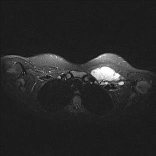
After the second biopsy which only confirmed lymphoproliferative disease, magnetic resonance imaging of the pectoral region revealed a mass of enlarged lymph nodes measuring 7.5 x 3.2 x 7 cm with the biggest lymph node measuring 6.1 cm in diameter (Figure 1).
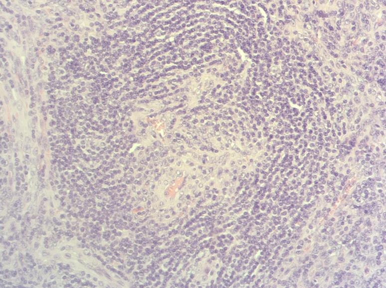
Due to the extent of local lymphadenopathy, neoadjuvant immunotherapy with 4 cycles of rituximab (375mg/m2 intravenously weekly) was administered. There was just a partial tumor reduction to neoadjuvant therapy, so the patient was reconsidered for surgery. Although blood tests disclosed preoperative anemia (Hb 10.8 g/L), there was no need for blood transfusions. The patient underwent a complete extirpation of the enlarged lymph nodes now measuring 4,2x2,7cm, 3,7x1,9 cm and 2,1x1,2 cm, respectively. Pathology finally confirmed Castleman disease of the multicentric hyaline‐vascular histologic type.
An interdisciplinary consultation was requested from the radiotherapist, who considered that adjuvant radiotherapy was not necessary. Two months after the operation, the patient made a full recovery without any complaints.
Three months after surgery, a high-resolution ultrasound examination demonstrated two enlarged lymph nodes inferior to the left subclavian vein. The patient underwent an MRI, which confirmed two large retropectoral lymph nodes in the clavicular region, measuring 3 and 2.5 cm.
We surgically removed the nodes, and the pathohistological report confirmed the previous diagnosis. Ultrasound examination was performed intraoperatively and showed no residual disease. One month after the surgery ultrasound follow-up showed one 2,2 cm enlarged lymph node below the acromial part of the left clavicle. We decided to “watch and wait” with regular ultrasound examinations. The patient remained asymptomatic, except for a temporary sensation of tension in the scar area during a three-year follow-up period. Finally, three years from the last excision, we excised the last enlarged lymph node, which measured 3.5 cm.
More than two years after the third surgical excision, the patient was still asymptomatic, without any lymphadenopathy, arm lymphedema or any abnormal laboratory findings.
3. Discussion
The major criteria to confirm idiopathic MCD are multicentric lymphadenopathy and biopsy-proven histopathology. The minor criteria are elevated CRP or erythrocyte sedimentation rate, anemia, thrombocytopenia or thrombocytosis, hypoalbuminemia, renal dysfunction, or proteinuria, polyclonal hypergammaglobulinemia, constitutional symptoms, hepatosplenomegaly, effusions or edema, cherry hemangiomata or violaceous papules, and lymphocytic interstitial pneumonitis [4]. Two of the minor criteria with at least one laboratory abnormality and exclusion of diseases that can mimic iMCD are required to confirm the diagnosis. Histologically, there are four different types: hyalin-vascular (HV), plasma-cell (PC), mixed and plasmablastic types [15,16].
The choice of therapy is still controversial in CD. Unlike the localized type, for which surgical excision is usually curative regardless of the histologic type, the multicentric disease generally requires aggressive systemic therapy but there are no uniform criteria [13,17].
In 2017, CDCN published the first guidelines for this disease [18]. So far, iMCD has been treated with a broad spectrum of agents, including corticosteroids, anti-CD 20 antibodies, cyclophosphamide, IFN, 2-chlorodeoxyadenosine, oral etoposide, vinblastine, liposomal doxorubicin, antibodies directed against IL-6 or IL-6 receptor and combination chemotherapy. All criteria (symptoms, laboratory results, and imaging) should be included when evaluating treatment response. Based on a review of 344 cases, the first evidence-based treatment guidelines for iMCD were established [19]. The working group recommends siltuximab, the anti-interleukin-6 monoclonal antibody, (11 mg/kg every three weeks) +/-corticosteroids as first-line therapy for all patients. Patients with a mild disease (without evidence of abnormal organ function that can require hospitalization but not in intensive care unit) who fail to respond to siltuximab after 3 to 4 doses adequately should receive rituximab (375 mg/m2 4-8 weekly doses) +/- corticosteroids +/- an immunomodulatory/ immunosuppressive agent, such as cyclosporine, sirolimus, anakinra, thalidomide, or bortezomib [4].
We treated the patient with rituximab since, at that time, siltuximab was not available. There is still a limited experience with anti-CD20 antibodies in treatment of patients diagnosed with CD. Recently, anti-CD20 monoclonal antibody (rituximab) is being used to treat HIV-positive MCD. There are two single case reports of treating HIV-negative patients with relapsed CD with rituximab along with cyclophosphamide and prednisone with favorable response [20,21]. According to Ide et al., rituximab might be also an appropriate first-line therapy for HIV-negative MCD patients. In their work they accomplished near a complete remission (66%) with no clinical symptoms due to MCD with a follow-up of 16-40 months after rituximab administration. Furthermore, they experienced no clinical remission of MCD after rituximab administration in one patient. These findings suggest that rituximab treatment might be as well an appropriate first-line therapy for HIV-negative MCD [22].
4. Conclusions
To sum up, localized and multicentric Castleman disease are different clinical disorders with overlapping histological features.
It seems that aggressive systemic therapy is indicated in the multicentric CD and may cure the disease with an often-favorable outcome. But according to our experience, our patient with diagnosed MCD stayed in remission without usage of aggressive therapy. Immunotherapy combined with surgical extirpation led to complete recovery in this 16-year-old patient.
In addition, the surgical procedure was not mutilated, which improved the quality of life for this young patient.
As in every rare disease, the recording of cases in rare disease registries is essential. Accumulation of the cases enables the evaluation of treatments and may provide a solid base for guidelines update.
Compliance with ethical standards
Any aspect of the work covered in this manuscript has been conducted with the ethical approval of all relevant bodies and that such approvals are acknowledged within the manuscript.
Abbreviations
- CD
-
Castleman disease
- MDC
-
Multicentric Castleman disease
- iMCD
-
idiopathic multicentric Castleman disease
- uCD
-
unicentric Castleman disease
- HV
-
hyalin vascular
- PB
-
plasma blastic
- CDCN
-
Castleman Disease Collaborative Network
- IFN
-
interferon
- IL
-
interleukin
References
- CASE records of the Massachusetts General Hospital Weekly Clinicopathological Exercises: Case 40011. N Engl J Med. 1954;250(1):26-30. doi: 10.1056/NEJM195401072500107
- Keller AR, Hochholzer L, Castleman B. Hyaline-vascular and plasma-cell types of giant lymph node hyperplasia of the mediastinum and other locations. Cancer. 1972;29(3):670-83. doi: 10.1002/1097-0142(197203)29:3<670::aid-cncr2820290321>3.0.co;2-#
- Munshi N, Mehra M, van de Velde H, Desai A, Potluri R, Vermeulen J. Use of a claims database to characterize and estimate the incidence rate for Castleman disease. Leuk Lymphoma. 2015;56(5):1252-60. doi: 10.3109/10428194.2014.953145
- Fajgenbaum DC. Novel insights and therapeutic approaches in idiopathic multicentric Castleman disease. Blood. 2018;132(22):2323-2330. doi: 10.1182/blood-2018-05-848671
- Cronin DM, Warnke RA. Castleman disease: an update on classification and the spectrum of associated lesions. Adv Anat Pathol. 2009;16(4):236-46. doi: 10.1097/PAP.0b013e3181a9d4d3
- Parez N, Bader-Meunier B, Roy CC, Dommergues JP. Paediatric Castleman disease: report of seven cases and review of the literature. Eur J Pediatr. 1999;158(8):631-7. doi: 10.1007/s004310051166
- Kligerman SJ, Auerbach A, Franks TJ, Galvin JR. Castleman Disease of the Thorax: Clinical, Radiologic, and Pathologic Correlation: From the Radiologic Pathology Archives. Radiographics. 2016;36(5):1309-32. doi: 10.1148/rg.2016160076
- Leger-Ravet MB, Peuchmaur M, Devergne O, Audouin J, Raphael M, Van Damme J, Galanaud P, Diebold J, Emilie D. Interleukin-6 gene expression in Castleman's disease. Blood. 1991;78(11):2923-30.
- Newlon JL, Couch M, Brennan J. Castleman's disease: three case reports and a review of the literature. Ear Nose Throat J. 2007;86(7):414-8.
- Roca B. Castleman's Disease. A Review. AIDS Rev. 2009;11(1):3-7.
- Szalat R, Munshi NC. Diagnosis of Castleman Disease. Hematol Oncol Clin North Am. 2018;32(1):53-64. doi: 10.1016/j.hoc.2017.09.005
- Wu D, Lim MS, Jaffe ES. Pathology of Castleman Disease. Hematol Oncol Clin North Am. 2018;32(1):37-52. doi: 10.1016/j.hoc.2017.09.004
- Oksenhendler E, Boutboul D, Fajgenbaum D, Mirouse A. The full spectrum of Castleman disease: 273 patients studied over 20 years. Br J Haematol. 2018;180(2):206-216. doi: 10.1111/bjh.15019
- McCarty MJ, Vukelja SJ, Banks PM, Weiss RB. Angiofollicular lymph node hyperplasia (Castleman's disease). Cancer Treat Rev. 1995;21(4):291-310. doi: 10.1016/0305-7372(95)90034-9
- Fajgenbaum DC, van Rhee F, Nabel CS. HHV-8-negative, idiopathic multicentric Castleman disease: novel insights into biology, pathogenesis, and therapy. Blood. 2014;123(19):2924-33. doi: 10.1182/blood-2013-12-545087
- Waterston A, Bower M. Fifty years of multicentric Castleman's disease. Acta Oncol. 2004;43(8):698-704. doi: 10.1080/02841860410002752
- Casper C. The aetiology and management of Castleman disease at 50 years: translating pathophysiology to patient care. Br J Haematol. 2005;129(1):3-17. doi: 10.1111/j.1365-2141.2004.05311.x
- Chronowski GM, Ha CS, Wilder RB, Cabanillas F, Manning J, Cox JD. Treatment of unicentric and multicentric Castleman disease and the role of radiotherapy. Cancer. 2001;92(3):670-6. doi: 10.1002/1097-0142(20010801)92:3<670::aid-cncr1369>3.0.co;2-q
- van Rhee F, Voorhees P, Dispenzieri A, Fosså A, Srkalovic G, Ide M, Munshi N, Schey S, Streetly M, Pierson SK, Partridge HL, Mukherjee S, Shilling D, Stone K, Greenway A, Ruth J, Lechowicz MJ, Chandrakasan S, Jayanthan R, Jaffe ES, Leitch H, Pemmaraju N, Chadburn A, Lim MS, Elenitoba-Johnson KS, Krymskaya V, Goodman A, Hoffmann C, Zinzani PL, Ferrero S, Terriou L, Sato Y, Simpson D, Wong R, Rossi JF, Nasta S, Yoshizaki K, Kurzrock R, Uldrick TS, Casper C, Oksenhendler E, Fajgenbaum DC. International, evidence-based consensus treatment guidelines for idiopathic multicentric Castleman disease. Blood. 2018;132(20):2115-2124. doi: 10.1182/blood-2018-07-862334
- Hudnall SD, Chen T, Brown K, Angel T, Schwartz MR, Tyring SK. Human herpesvirus-8-positive microvenular hemangioma in POEMS syndrome. Arch Pathol Lab Med. 2003;127(8):1034-6. doi: 10.5858/2003-127-1034-HHMHIP
- Abdou S, Salib H. An extra ordinary response of Castleman’s disease to rituximab. Blood. 2004;104:49b-49b.
- Ide M, Kawachi Y, Izumi Y. Long-term remission in HIV-negative patients with multicentric Castleman's disease using rituximab. Eur J Haematol. 2006;76(2):119-23. doi: 10.1111/j.1600-0609.2005.00570.x
