

Acute parotiditis after cesarean section; case report
1 St. John Hospital, Bucur Maternity, Department of Obstetrics and Gynecology, Bucharest, Romania
2 Carol Davila University, Department of Obstetrics and Gynecology, Bucharest, Romania
Abstract
The enlargement of the parotid gland develops in inflammatory or stenotic conditions but after Cesarean section the symptomatology is unusual. A 38 year old patient with no obstetrical history referred to our clinic for pregnancy, which followed our national program of prenatal care. The outcome of the pregnancy was favorable for both mother and fetus. During labor the fetus developed bradycardia and the patient delivered by Cesarean section a 3400 g baby-boy with 8 Apgar Score; the anesthesia was spinal.
18 hours after delivery the patient presented mild respiratory distress. The symptomatology was caused by the enlargement of the parotid gland. The treatment was supportive and the remission occurred 10 hours after the onset. The initial discussion that raised this case was caused by the viral, infection and stenotic cause of the parotiditis. All these reasons had no medical argument. It was also debated about the anesthesia but, until now, the medical literature didn’t report any case of association between parotiditis and spinal anesthesia. It is also impossible to correlate the parotiditis with IVF procedure.
As a conclusion, this case is unique because it represents the parotiditis without unknown case that appeared after Cesarean section and spinal anesthesia in a healthy woman.
1. Introduction
The parotid gland is the largest of the major salivary glands. The parotid glands are situated on the anterior site of the external auditory canal, inferior to the zygomatic arch and superior to the mandibular angle. The salivary gland has 20 to 30 lymph nodes. The lymphatic drainage is from the ipsilateral side of the face and forehead, the auricular region and the external auditory canal [1]. The enlargement of the parotid gland develops in inflammatory or stenotic conditions but after Cesarean section the symptomatology is unusual.
Case presentation
The patient CM, 38 years of age, referred to our clinic for pregnancy prenatal care. She had no obstetrical history (other pregnancy or abortion before). Due to poor ovarian reserve (Antimullerian Hormone 0.01) she obtained this pregnancy using ART procedures.
The patient underwent in vitro fertilization. The procedure had no complication. The pregnancy followed our national program of prenatal care. The biological findings were normal in the first trimester of pregnancy (hemoglobin lever, red blood count, renal and hepatic function evaluation, blood sugar level, urinary evaluation, etc.). The first trimester ultrasound examination revealed no abnormal findings. The first trimester evaluation for trisomy 21, 13 and 18 was negative (combined test NT 2,4 mm and present nasal bone, no tricuspid valve regurgitation). The second trimester ultrasound scan also revealed a normal fetal anatomy. The outcome of the pregnancy was favorable for both mother and fetus. In the third trimester of pregnancy the obstetrical ultrasound revealed nuchal cord. During labor the fetus developed bradycardia and the patient delivered by Cesarean section a 3400 g baby-boy with 8 Apgar Score. The anesthesia was spinal. The fetal outcome was favorable.
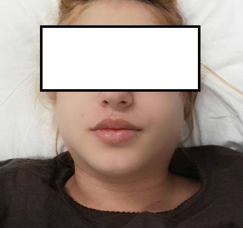
The peculiarity of this case is the early postoperative maternal outcome. 18 hours after delivery the patient presented mild respiratory distress. The respiratory symptomatology was caused by the edema of the anterior cervical region. The clinical examination revealed bilateral enlarged parotid gland (Figure 1). The patient received antihistaminic and anti-inflammatory drugs to prevent glotic edema. The ENT examination confirmed the bilateral parotiditis but it couldn't reveal the cause. Antiurlian virus antibodies IgM were determined and were negative while IgG were positive excluding retrospectively an urlian ethiology of the parotid inflamation. The treatment was supportive and the remission occurred 10 hours after the onset. The initial discussion that raised this case was caused by the viral, infection and stenotic cause of the parotiditis. All these reasons had no argument. It was also debated about the anesthesia but, until now, the medical literature didn’t report any case of association with between parotiditis and spinal anesthesia. It is also impossible to correlate the parotiditis with IVF procedure.
2. Discussion
The parotid duct, named the Stensen's duct, is the main excretory duct for parotid gland. Saliva from the parotid gland flows through the Stenon duct into the oral cavity in the region of the second maxillary molar. The mechanism of disrupting salivary flow through the parotid duct, such as anatomic anomaly, mass, sialolithiasis, stenosis or medications can cause parotiditis. Some of these obstruction can determine bacterial (acute suppurative parotiditis), viral (mumps), inflammatory (due to obstruction such as stone or duct stricture) and neoplastic complications [2, 3]. If salivary stasis occurs then the retrograde seeding of the Stensen's duct by a mixed oral flora acute bacterial suppurative parotiditis may develop [4]. If the embryogenic cysts infects, such as the first branchial cleft, suppuration of the parotid gland may also occur [5].
It was described that in the postoperative period suppurative parotiditis may occur in elderly patients who are intubated or dehydrated, but it may also be seen in outpatients [6]. There were identified predisposing factors that include recent intensive teeth cleaning, salivary calculi with obstruction, malnutrition, neoplasm of the oral cavity anticholinergic drugs and drugs that reduce salivary flow [7].
A recent critical review of this topic concluded that only three drugs have been suggested as associated with the acute parotiditis: l-asparaginase, clozapine, and phenylbutazone. They have sufficient evidence to cause a direct relationship of ductus obstruction [8]. General anesthesia has long been identified as a very rare cause of acute parotiditis known as “anesthesia mumps”. Even if this entity is generally bilateral [9, 10], there were some cases where it has been reported with unilateral symptomatology [11]. Moreover noinvasive critical care respiratory ventilation was described as causally linked to the development of acute parotiditis. It is due to transmission of positive pressure to the oral cavity causing obstruction of salivary flow through the parotid duct. Clinicians should be able to recognize this possible complication of ventilation [12].
Many medical reports have suggested different methods to prevent the development of anesthesia mumps. There were proposed adaptive-shaped soft pads conservatively and changing the head and neck position to avoid mechanical occlusion of the parotid gland and ducts [13]. According to this condition, careful consideration should be given to patients with predisposing conditions like a long operation time, premedication involving anticholinergics, prone position, obesity, and a history of parotid disease or trauma. In addition, anesthesiologists should be aware that anesthesia mumps can develop with usual face mask handling, and a gentle procedure is always required [14].
3. Conclusions
The presented case is special because it represents the parotiditis without unknown etiology that appeared after Cesarean section and spinal anesthesia in a healthy woman.
References
- Tan VE, Goh BS. Parotid abscess: a five-year review--clinical presentation, diagnosis and management. J Laryngol Otol. 2007; 121(9): 872- 9. PMID: 17112392, https://doi.org/10.1017/S0022215106004166
- Hernandez S, Busso C, Walvekar RR. Parotitis and Sialendoscopy of the Parotid Gland. Otolaryngol Clin North Am. 2016; 49(2): 381-93. PMID: 26912292, https://doi.org/10.1016/j.otc.2015.12.003
- Capaccio P, Gaffuri M, Pignataro L, Assandri F, Pereira P, Farronato G. Recurrent parotid swelling secondary to masseter muscle hypertrophy: a multidisciplinary diagnostic and therapeutic approach. Cranio. 2016; 34(6): 388-94. PMID: 26892838, https://doi.org/10.1080/08869634.2016.1142694
- Cohen MA, Docktor JW. Acute suppurative parotitis with spread to the deep neck spaces. Am J Emerg Med. 1999; 17(1): 46-9. PMID: 9928699, https://doi.org/10.1016/S0735-6757(99)90015-3
- Triglia JM, Nicollas R, Ducroz V, Koltai PJ, Garabedian EN. First branchial cleft anomalies: a study of 39 cases and a review of the literature. Arch Otolaryngol Head Neck Surg. 1998; 124(3): 291-5. PMID: 9525513, https://doi.org/10.1001/archotol.124.3.291
- Brook I. The swollen neck. Cervical lymphadenitis, parotitis, thyroiditis, and infected cysts. Infect Dis Clin North Am. 1988; 2(1): 221- 36. PMID: 3074109
- Hamid R, Shera HA, Khurshid S, Bhat AN, Baba AA, Akhter A. Suppurative parotitis and submandibular sialadenitis. J Clin Neonatol. 2014; 3(1): 39-40. PMID: 24741539, https://doi.org/10.4103/2249-4847.128730
- Brooks KG, Thompson DF. A review and assessment of drug-induced parotitis. Ann Pharmacother. 2012; 46(12): 1688-99. PMID: 23249870, https://doi.org/10.1345/aph.1R228
- Reilly DJ. Benign transient swelling of the parotid glands following general anesthesia: “anesthesia mumps Anesth Analg. 1970; 49(4): 560-3. PMID: 5534667, https://doi.org/10.1213/00000539- 197007000-00009
- Jafra A, Arora S, Dwivedi D. Benign swelling of submandibular glands under general anesthesia "anesthesia mumps". J Clin Anesth. 2016; 34: 325- 6. PMID: 27687402, https://doi.org/10.1016/j.jclinane.2016.05.016
- Kwon SY, Kang YJ, Seo KH, Kim Y. Acute unilateral anesthesia mumps after hysteroscopic surgery under general anesthesia: a case report. Korean J Anesthesiol. 2015; 68(3): 300-3. PMID: 26045936, https://doi.org/10.4097/kjae.2015.68.3.300
- Martinez E, Dicpinigaitis PV. Development of acute parotitis after non-invasive ventilation. J Thorac Dis. 2017; 9(7): E605-E608. PMID: 28840025, https://doi.org/10.21037/jtd.2017.06.86
- Bahadur S, Fayyaz M, Mehboob S. Salivary gland swelling developing after endoscopy: "anesthesia mumps". Gastrointest Endosc. 2006; 63(2): 345–7. PMID: 16427956, https://doi.org/10.1016/j.gie.2005.09.008
- Tian X, Eldadah M, Cheng W. Neonatal Suppurative Parotitis: Two Cases. Pediatr Infect Dis J. 2016; 35(7): 823-4. PMID: 27351362, https://doi.org/10.1097/INF.0000000000001177
