

Transduodenal Local Resection of Ampullary Neuroendocrine Tumors with Bleeding
1 Health Sciences University, Istanbul Fatih Sultan Mehmet Education nad Research Hospital, Department of General Surgery, Istanbul, Turkey (34734)
Abstract
Duodenal neuroendocrine tumors in the Ampulla of Vater occur very rarely and are very difficult to diagnose preoperatively. Duodenal ulcer bleeding due to the destruction of the duodenal mucosa is very rare. In this study, we present the case of a duodenal neuroendocrine tumor presented with upper gastrointestinal bleeding, which was treated by means of transduodenal local resection.
As a conclusion, endoscopic or transduodenal local excision is relatively safe to be used in ampullary neuroendocrine tumors with no distant metastasis or local invasions. Endoscopic resection is recommended in patients with low grade tumors within the submucosa, smaller than 2 cm and with a low KI-67 index. In support of this, EUS has been adopted as an important tool to measure the depth of invasion and evaluate the lymph node status in staging the gastrointestinal tumors as well as collecting specimens simultaneously.
Finally, endoscopic procedures (ESD - EMR) present a higher risk of perforation, so that a large number of prospective controlled studies are needed to establish a consensus on this therapeutic approach.
Introduction
The small bowel neuroendocrine tumors are the most common subgroup among the gastrointestinal neuroendocrine tumors. The incidence of a small intestinal neuroendocrine tumor is continuously increasing [1]. Neuroendocrine tumors constitute approximately 2% of all types of neuroendocrine tumors. Duodenal neuroendocrine tumors constitute 5% of all gastrointestinal neuroendocrine tumors. It is usually detected in the form of solitary small masses located in the duodenal submucosa. Only 0.3% of all neuroendocrine tumors are located around the Ampulla of Vater if we take into account the gastrointestinal tract [2]. However, the rate rises as the incidence and prevalence of upper gastrointestinal endoscopy increase.
These tumors are categorized into functional and non-functional. The functional group consists of 27-58% gastrinoma, 23-75% somatostatinoma, 28% serotonin-secreting tumors, 9% calcitonin secreting tumors and rarely gangliocytic paragangliomas and non-functional tumors [3]. According to WHO 2010 classification, duodenal neuroendocrine tumors are graded as follows: grade G1 (Ki-67 B 2%), G2 (Ki-67 3-20%), G3 (Ki-67 20%) [4]. Symptoms of periampullary neuroendocrine tumors mostly depend on their location: pain (37%), jaundice (18%), nausea/vomiting (4%), bleeding (2%), anemia (21%), diarrhea (4%) and duodenal obstruction (1%) [5]. Duodenal periampullary neuroendocrine tumors occur very rarely. As a result, there is no current definitive approach in the treatment of these tumors. A wide range of treatment options has been reported in the literature, from endoscopic procedures to a radical surgical method, i.e. pancreaticoduodenectomy [6].
Prognosis may vary according to the histopathological type of the tumor. It is categorized into 3 different histopathological groups as follows: [1] well-differentiated neuroendocrine tumor (carcinoid), [2] well-differentiated neuroendocrine carcinoma (malignant carcinoid), [3] poorly differentiated neuroendocrine carcinoma (malignant carcinoid) [1].
There is not enough information about the prognostic factors due to the limited data in the literature. In this case report, we present a patient diagnosed with neuroendocrine tumors of the Ampulla of Vater with upper gastrointestinal bleeding, who was treated by means of transduodenal local resection [7].
Case report
A 67-year-old female patient was admitted to the emergency department with the complaint of 1-week history of melena. The patient's medical history included the diagnosis of hypertension. On physical examination, the patient showed hemodynamic instability, arterial blood pressure of 90/60 mmHg and a heart rate of 110/min along with sinus tachycardia. The presence of melena was detected on rectal examination. The hemoglobin level was 7 g/dL. Bilirubin, liver enzymes, prothrombin time and tumor markers in the blood were normal. The replacement of 2 units of erythrocyte suspension was performed due to the hemodynamic instability. Emergency upper gastrointestinal endoscopy was performed after the patient became stable. During endoscopy, a 3-cm ulcerative mass was detected around the duodenal papilla in the middle of the second and third portions of the duodenum (Figure 1).
No active bleeding was detected during endoscopy and multiple samples were taken from the sites around the ulcer. The histopathology of endoscopic biopsies revealed chronic duodenitis. Magnetic resonance imaging (MRI) revealed a different type of mass of 2.5x3.5 cm in the duodenal wall, in the second portion of the duodenum. MRI demonstrated minimal dilatation of the intrahepatic bile ducts and the pancreatic duct (Figure 2). No local invasion, regional lymph nodes nor distant metastasis were detected on MRI. Upper Gastrointestinal Endosonography (EUS) was planned in order to evaluate the condition of the hepatobiliary system, pancreatic duct, duodenal papilla and duodenal wall. EUS revealed a heterogeneous mass of 3.5x2.5 cm with hypoechoic and calcified areas in the duodenal mucosa. The mass was detected to be associated with the Ampulla of Vater. No pathology was detected in the pancreas, gallbladder and biliary tract by EUS.
The patient was informed and gave his consent for both methods of transduodenal resection and pancreatico-duodenectomy. Kocher maneuver was performed following the median incision. In the second portion of the duodenum, the tumor presented as palpable and mobile. A 5-cm polypoid mass was detected with overlying mucosal ulceration after duodenotomy (Figures 3-4).
The transduodenal resection was performed followed by cholecystectomy and ampullary sphincteroplasty after resection. The margins were sent for the frozen section and returned negative for the gastrointestinal stromal tumor. The cholangiography of the cystic duct during surgery revealed normal bile ducts and pancreatic ducts.
The pathological macro and microscopic examination of the excised material revealed epithelioid and circular cell proliferation exhibiting a solid growth pattern. In the immune-histochemical examination for KI-67, the result was 1%. Tumor S-100, chromogranin and synaptophysin were positive, while Desmin, CD 34 and C-KIT were negative. Finally, the duodenal papilla was diagnosed as well-differentiated neuroendocrine carcinoma (pT2 NX M0) (Figure 5).


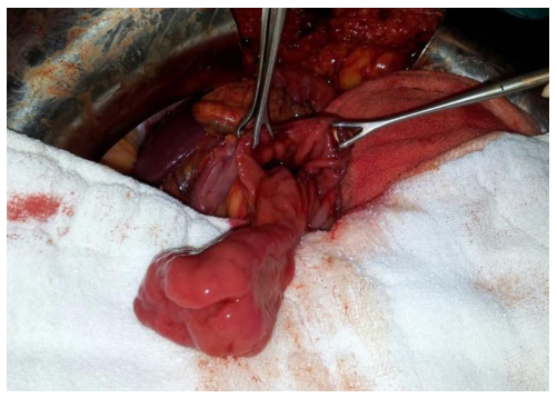

surgery
There were no complications in the postoperative period and the patient was discharged on the 5th postoperative day. A whole-body octreotide scintigraphy was performed following the definitive diagnosis of neuroendocrine carcinoma and no metastatic focus was identified (Figure 6). The follow-up of the patient a year later revealed no signs of recurrence.

Discussions
The recent increase in the number of neuroendocrine tumors of the duodenum is due to the increase in cancer screening and applications of endoscopy. Diagnosis is usually made by means of upper gastrointestinal endoscopy and staging is completed by means of computed tomography [2, 7]. EUS, which allows local and regional applications, can also be used for diagnosis. The prognosis of duodenal neuroendocrine tumors varies greatly due to the advanced heterogeneity, therefore, the treatment options should be evaluated exclusively for each patient. Research reveals that the majority of duodenal neuroendocrine tumors are made up of localized tumors and are smaller than 2 cm. Only 10% are regional and 9% indicate metastatic diseases [8].
The factors indicating metastatic disease are: a lesion greater than 1 cm, the extension of the muscularis propria being slightly differentiated and the lymphovascular invasion [9].
The most common clinical manifestation of duodenal neuroendocrine tumors is usually dyspepsia. In addition, pain, jaundice, nausea and vomiting, anemia, bleeding, diarrhea and duodenal obstruction may also be present. Symptoms such as jaundice, biliary duct dilatation, nausea, vomiting and diarrhea are more common in the neuroendocrine tumors of the Ampulla of Vater. Periampullary neuroendocrine tumors are frequently associated with Von Recklinghausen Disease (18%). Chromogranin A should be considered in case of suspicion of a duodenal neuroendocrine tumor as 60 to 100% of the patients’ test results are positive. In a retrospective study of 1914 patients with gastrointestinal neuroendocrine tumors, the patients were examined for the size of the tumors limited to the submucosa and the results revealed that the tumor was smaller than 0.5 cm in 8.3% of cases, smaller than 1 cm in 10.5% of cases, smaller than 2 cm in 13.8% of cases and bigger than 2 cm in 25.8% of cases. The same study examined the depth of invasion of the tumor and revealed that 1.7% of the tumors were mucosal, 10.5% were submucosal, 29.6% were in the muscularis propria and 42.8% were serosal or subserosal. The risk of metastasis is likely to increase as the size of the gastrointestinal neuroendocrine tumors increases [9].
Since the studies in the literature are retrospective, our knowledge is limited, with questionable reliability. Pancreaticoduodenectomy has advantages as it allows both the complete removal of the tumor and the detailed examination of the surrounding tissue and lymph nodes. However, it has higher mortality and morbity rates compared to other methods. In the study conducted by Bobby et al., it was stated that panreaticoduodenectomy should be performed in lesions larger than 2 cm due to its surgical margin (R0) and low recurrence rates. A 20% lymph node metastasis was detected in the patients who underwent pancreaticoduodenectomy, whereas the recurrence should be higher in patients who did not undergo lymph node dissection [10]. Burke et al. found that the rate of lymph node metastasis detected during pancreaticoduodenectomy was above 50%, but did not lead to changes in life expectancy. Since duodenal neuroendocrine tumors have a high rate of lymph node metastasis, an accurate staging should be performed prior to any kind of operation or endoscopy [11].
The presence of distant metastases should be excluded preoperatively. The follow-up of metastasis and local recurrence should be done regularly due to the heterogeneity of the disease and various prognostic features. Some other evidence is that the size of the ampullary and periampullary lesions is greater than the others and, having a higher grade, the metastasis rates are also higher [12].
Pancreaticoduodenectomy has higher mortality and morbidity rates compared to the others, but it was noted that when it was performed in specialized high-volume centers, the postoperative quality of life was not lower than that of patients undergoing different methods of treatment. KI-67 status and the grading system, which are considered to be critical prognostic factors, have not been sufficiently used in current studies [13, 14].
EUS has been adopted as an important tool to measure the depth of invasion and evaluate the lymph node status in staging the gastrointestinal tumors as well as collecting specimens simultaneously [8].
Endoscopic resection is recommended in patients with low grade tumors within the submucosa, smaller than 2 cm and with a low KI-67 index. Pancreaticoduodenectomy at a high-volume center should be opted for in cases with high grade and KI-67 index, lesion greater than 2 cm, the invasion of the muscularis propria and lymph node metastasis providing that the patient is capable of coping with radical surgery [13, 14]. Although the duodenal neuroendocrine tumors resemble the gastric neuroendocrine tumors in terms of being well-differentiated and being low-grade, they are not as suitable for endoscopic interventions as the stomach due to the relatively thin duodenal wall. Endoscopic procedures (ESD - EMR) have a higher risk of perforation. A large number of prospective controlled studies are needed to establish a consensus on this issue.
Conclusions
The neuroendocrine tumors of the Ampulla of Vater occur very rarely. There are several cases in the literature diagnosed by means of emergency endoscopy following upper gastrointestinal bleeding. Endoscopic or transduodenal local excision is a safe method to use in ampullary neuroendocrine tumors with no distant metastasis or local invasion.
Institutional Review Board Statement
Any aspect of the work covered in this manuscript has been conducted with the ethical approval of all relevant bodies and that such approvals are acknowledged within the manuscript.
Conflicts of Interest
There are no known conflicts of interest in the publication of this article. The manuscript was read and approved by all authors.
References
- Fitzgerald TL, Dennis SO, Kachare SD, et al. Increasing incidence of duodenal neuroendocrine tumors: incidental discovery of indolent disease? Surgery. 2015; 158(2): 466–71. DOI: 10.1016/j.surg.2015.03.042
- Yao JC, Hassan M, Phan A, Dagohoy C, Leary C, Mares JE, et al. One Hundred Years After “Carcinoid”: Epidemiology of and Prognostic Factors for Neuroendocrine Tumors in 35,825 Case in the United States. J Clin Oncol. 2008; 26(18): 3063–72. PMID: 18565894, DOI: 10.1200/JCO.2007.15.4377
- Jensen RT, Rindi G, Arnold R, et al. Frascati Consensus Conference; European Neuroendocrine Tumor Society. Well differentiated duodenal tumor/ carcinoma (excluding gastrinomas). Neuroendocrinology. 2006; 84(3): 165–72. PMID: 17312376 DOI: 10.1159/000098008
- Rindi G, Klo¨ppel G, Alhman H, et al.; all other Frascati Consensus Conference participants; European Neuroendocrine Tumor Society (ENETS); European Neuroendocrine Tumor Society (ENETS). TNM staging of foregut (neuro)endocrine tumors: a consensus proposal including a grading system. Virchows Arch. 2006; 449(4): 395–401. PMID: 16967267, DOI: 10.1007/s00428-006-0250-1
- Delle Fave G, O’Toole D, Sundin A, Taal B, Ferolla P, Ramage JK, Ferone D, Ito T, Weber W, Zheng-Pei Z, De Herder WW, Pascher A, Ruszniewski P; Vienna Consensus Conference participants. ENETS consensus guidelinesupdate for gastroduodenal neuroendocrine neoplasms. Neuroendocrinology. 2016; 103(2): 119– 24. PMID: 26784901, DOI: 10.1159/000443168
- Shroff SR, Kushnir VM, Wani SB, Gupta N, Jonnalagadda SS, Murad F, et al. Efficacy of Endoscopic Mucosal Resection for Management of Small Duodenal Neuroendocrine Tumors. Surg Laparosc Endosc Percutan Tech. 2015; 25(5): e134–9. PMID: 26271024, DOI: 10.1097/SLE.0000000000000192
- Kwon YH, Jeon SW, Kim GH, Kim JI, Chung IK, Jee SR, KimHU, Seo GS, Baik GH, Choi KD, Moon JS. Long-term follow up of endoscopic resection for type 3 gastric NET. World J Gastroenterol. 2013; 19(46): 8703-8. PMID: 24379589, DOI: 10.3748/wjg.v19.i46.8703
- HoffmannKM, FurukawaM, Jensen RT. Duodenal neuroendocrine tumors: Classification, functional syndromes, diagnosis and medicaltreatment. Best Pract Res Clin Gastroenterol. 2005; 19(5): 675–97. PMID: 16253893, DOI: 10.1016/j.bpg.2005.05.009
- Soga J. Early-stage carcinoids of the gastrointestinal tract: an analysisof 1914 reported cases. Cancer. 2005; 103(8): 1587–95. PMID: 15742328, DOI: 10.1002/cncr.20939
- Dasari BVM, Al-Shakhshir S, Pawlik TM, Shah T, Marudanayagam R, Sutcliffe RP, Mirza DF, Muiesan P, Roberts KJ, Isaac J2. Outcomes of Surgical and Endoscopic Resection of Duodenal Neuroendocrine Tumours (NETs): a Systematic Review of the Literature. J Gastrointest Surg. 2018; 22(9): 1652-8. PMID: 29869091, DOI: 10.1007/s11605-018-3825-7
- Burke CA, Beck GJ, Church JM, van Stolk RU. The natural history of untreated duodenal and ampullary adenomas in patients with familial adenomatous polyposis followed in an endoscopic surveillance program. Gastrointest Endosc. 1999; 49(3 Pt 1): 358– 64. PMID: 10049420
- Witzigmann H, Loracher C, Geissler F, Wagner T, Tannapfel A,Uhlmann D, et al. Neuroendocrine tumours of the duodenum: Clinical aspects, pathomorphology and therapy. Langenbeck’s Arch Surg. 2002; 386(7): 525–33. PMID: 11819111, DOI: 10.1007/s00423-001-0260-z
- Min B-H, Kim ER, Lee JH, Kim K-M, Min YW, Rhee P-L, Kim JJ, Rhee JC. Management Strategy for Small Duodenal Carcinoid Tumors: Does Conservative Management with Close Follow-Up Represent an Alternative to Endoscopic Treatment. Digestion. 2013; 87(4): 247–53. PMID: 23751414, DOI: 10.1159/000349958
- Delle Fave G, Kwekkeboom DJ, Van Cutsem E, Rindi G, Kos-Kudla B, Knigge U, Sasano H, Tomassetti P, Salazar R, Ruszniewski P; Barcelona Consensus Conference participants. ENETS Consensus Guidelines for the Management of Patients with Gastroduodenal Neoplasms. Neuroendocrinology. 2012; 95(2): 74–87. PMID: 22262004, DOI: 10.1159/000335595
