

A case of ectopic fibroadenoma mimicking left axillary lymphadenopathy
1 VOCATIONAL SCHOOL OF HEALTH SERVICES, ISTANBUL RUMELI UNIVERSITY, DEPARTMENT OF MEDICAL IMAGING TECHNIQUES, ISTANBUL, TURKEY
2 ACIBADEM KOZYATAGI HOSPITAL, DEPARTMENT OF RADIOLOGY, ISTANBUL, TURKEY
Abstract
Fibroadenomas are the most common benign tumors of the breast. Since they originate from the fibroglandular breast tissue, they cannot be localized anywhere outside the breast. However, they can be localized in accessory or ectopic breast tissue and cause confusion in terms of clinical differential diagnosis. Small ectopic fibroadenomas placed in normal breast tissue are generally asymptomatic and nonpalpable. In contrast, lesions located in the axillary region are usually palpable, due to their superficial location and skin that is thinner at this level. This case report presents the clinical and imaging findings of a patient with a preliminary diagnosis of lymphadenopathy, with a superficial axillary location of the lesion and who underwent biopsy. In our case, no obvious accessory breast tissue was detected by physical examination or ultrasound of the left armpit. Even though there was axillary accessory breast tissue, it was not at a level that could be detected by ultrasound.
Introduction
Developmental anomalies of the breast are rare conditions. The most common developmental anomaly of the breast is the accessory breast, with a rate of 2-6% [1]. Fibroadenomas, the most common solid tumor of the breast, can also develop from these accessory breast tissues. While small-sized ectopic fibroadenomas in normal breast tissue are usually asymptomatic and nonpalpable, lesions located in the axillary region are usually palpable due to more superficial localization and thinner skin at this level [2]. In addition, it can be large and painful in the premenstrual period due to the influence of menstrual cyclic hormonal changes, and patients may present for this reason [3]. In this case report are presented clinical and imaging features of a 40-year-old female patient, who presented with a sudden onset of painless hard mass in her left axilla and was diagnosed with fibroadenoma histopathologically.
Case report
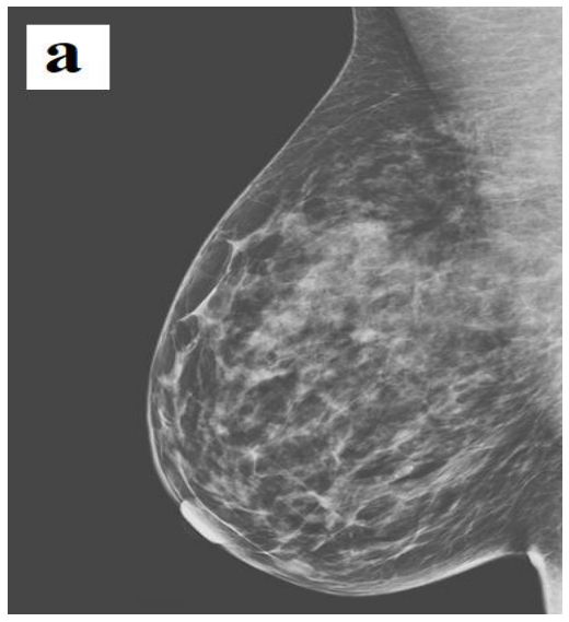
A 40-year-old female patient was admitted to our hospital due to newly developed and palpable painless stiffness in the left axilla. At the physical examination of the patient, no palpable lesion was detected in both breasts. In the axillary examination, palpable lymphadenopathy was not detected, except for mobile stiffness with smooth contours noticed by the patient at the level of the anterior axillary line in the left axilla. There was no physical examination finding of accessory breast tissue. The medical history of the case found that there was no comorbid disease and no family history in terms of breast cancer. The newly developed left axillary lesion detected on physical examination was evaluated in favor of lymphadenopathy, and the case was referred for ultrasonographic and mammographic evaluation. Dense breast structure and bilateral axillary lymph nodes were detected in both breasts on mammography. Palpable stiffness in the left axilla was also evident in the mammographic examination as a nodular opacity with smooth contours in the upper axillary region (Figure 1). No space-occupying solid-cystic lesion was detected in both breasts in ultrasonography.
In the left axilla, a well-contoured, oval-shaped, homogeneous internal structure, hypovascularized 13x5 mm solid lesion was detected by color Doppler
Deniz Esin Tekcan Sanli sonography, and hilar vascular structures could not be detected (Figure 2).
It was evaluated in favor of pathological LAP, and control was recommended after antibiotic treatment. The lesion described was stable in the sonographic examination of the patient who came to the follow-up at the 1st month after amoxicillin 1000 mg (2x1) treatment. No significant regression was found in terms of size. No new formation was detected in both breasts and axillae. The histopathological diagnosis of the lesion, which was sampled with tru-cut biopsy in terms of differential diagnosis, was fibroadenoma (Figure 3). The case was followed up with the diagnosis of left axillary ectopic fibroadenoma. Written informed consent was obtained from the patient.


Discussion
Fibroadenomas are the most common lesions of the breast after cysts. Since they are composed of fibroglandular breast tissue components, just like the breast tissue, their internal structures may change depending on hormonal factors and may show some degeneration patterns [4-6]. They can also be confused with malignancy in terms of radiological imaging features if degenerated or rapidly progressive. It is very rare for them to be located elsewhere than in the breast tissue [2,7]. In a few examples reported in the literature, the most common ectopic location is the axillary region, where the accessory breast tissue is most common. These ectopic and atypical locations may also bring to mind the possibility of a possible malignant lesion.
In our case, no obvious accessory breast tissue was detected sonographically and by physical examination in the left axilla. Therefore, the lesion was initially evaluated in favor of LAP. The reason for this can be explained by the fact that the patient is very thin and the breast tissue is noticeably small. Even if there is axillary accessory breast tissue, it is not at a level that can be detected sonographically.
Therefore, even if there is no obvious accessory breast tissue, the possibility of ectopic fibroadenoma should be kept in mind in the differential diagnosis of axillary LAPs whose hilum is not selected. Moreover, echogenic septations considered characteristic for fibroadenomas can sometimes be confused with the hilum of the lymph node. In this case, the thing to do is to investigate vascularization with color Doppler sonography. While vascular structures can usually be found in the hilum of lymph nodes, echogenic septations of fibroadenomas are hypo-avascular [8,9]. In addition, some degenerative changes may occur in fibroadenomas with some hormonal changes and they may increase in size rapidly during pregnancy-lactation periods. Therefore, whether localized in the breast or ectopic, they should be investigated for malignancy. Radiological imaging methods are not always successful in making this distinction. Therefore, histopathological sampling is often needed.
There are also a few interestingly reported cases of vulvar fibroadenoma other than the axilla [10,11]. Therefore, it should be kept in mind that ectopic fibroadenomas may be present in the differential diagnosis of sonographically smooth-contoured, oval-shaped, hypovascularized lesions in any atypical localizations. In addition, it should not be forgotten that ectopic fibroadenoma can develop wherever there is ectopic breast tissue. In cases reported so far, it has been shown that accessory breast tissue can be localized on the back, face, neck, shoulder, arm, and hip [12-14]. When a solid lesion is encountered in such atypical localizations, ectopic breast tissue should be considered when making the differential diagnosis.
Conclusions
In conclusion, although ectopic fibroadenomas are generally benign conditions, they must be confirmed histopathologically. It is a consequence of the fact that clinical and imaging data may suggest malignant processes and pathological LAPs, especially in elderly cases and degenerated fibroadenomas.
Institutional Review Board Statement
Any aspect of the work covered in this manuscript has been conducted with the ethical approval of all relevant bodies and that such approvals are acknowledged within the manuscript.
Conflicts of Interest
There are no known conflicts of interest in the publication of this article. The manuscript was read and approved by all authors.
References
- Mukhopadhyay M, Saha AK, Sarkar A. Fibroadenoma of the ectopic breast of the axilla. Indian J Surg. 2010 Apr;72(2):143-5. doi: 10.1007/s12262-010-0024-5
- Ravikanth R, Majumdar P. Fibroadenoma in Ectopic Breast Tissue of Axilla: A Rare Entity. J Med Ultrasound. 2019;28(1):50-51. doi: 10.4103/JMU.JMU_65_19
- Goyal S, Bawa R, Sangwan S, Singh P. Fibroadenoma of axillary ectopic breast tissue: A rare clinical entity. Clin Cancer Investig J. 2014;3(3):242-4.
- Littrup PJ, Duric N, Sak M, Li C, Roy O, Brem RF, Larsen LH, Yamashita M. Multicenter Study of Whole Breast Stiffness Imaging by Ultrasound Tomography (SoftVue) for Characterization of Breast Tissues and Masses. J Clin Med. 2021 Nov 25;10(23):5528. doi: 10.3390/jcm10235528
- Moini A, Eslami B, Alipour S. Breast fibroadenomas are less frequent in women with uterine fibroids. Breast Dis. 2022;41(1):51-54. doi: 10.3233/BD-210009
- Johansson A, Christakou AE, Iftimi A, Eriksson M, Tapia J, Skoog L, Benz CC, Rodriguez-Wallberg KA, Hall P, Czene K, Lindström LS. Characterization of Benign Breast Diseases and Association With Age, Hormonal Factors, and Family History of Breast Cancer Among Women in Sweden. JAMA Netw Open. 2021;4(6): e2114716. doi: 10.1001/jamanetworkopen.2021.14716
- Yalagachin G, Lakshmikantha N, Mashal SB. Prevalence of nodular goiter in patients with breast diseases. J Clin Invest Surg. 2020;5(2):91-95. doi: 10.25083/2559.5555/5.2/91.95
- Grandl S, Willner M, Herzen J, Sztrókay-Gaul A, Mayr D, Auweter SD, Hipp A, Birnbacher L, Marschner M, Chabior M, Reiser M, Pfeiffer F, Bamberg F, Hellerhoff K. Visualizing typical features of breast fibroadenomas using phase-contrast CT: an ex-vivo study. PLoS One. 2014 May 13;9(5):e97101. doi: 10.1371/journal.pone.0097101
- Balalau OD, Scaunasu RV, Olaru OG, Dumitriu AS, et al. Modern interpretation of risk factors in breast cancer of women. J Mind Med Sci. 2022;9(1):88-95. doi: 10.22543/7674.91.P8895 Deniz Esin Tekcan Sanli
- Kalyani R, Srinivas MV, Veda P. Vulval fibroadenoma - a report of two cases with review of literature. Int J Biomed Sci. 2014 Jun;10(2):143-5.
- Lev-Cohain N, Kapur P, Pedrosa I. Vulvar fibroadenoma with lactational changes in ectopic breast tissue. Case Rep Obstet Gynecol. 2013;2013:924902. doi: 10.1155/2013/924902
- Tiwary SK, Kumar P, Khanna AK. Fibroadenoma in axilla: another manifestation of ectopic breast. BMJ Case Rep. 2015 Apr 26;2015:bcr2015209535. doi: 10.1136/bcr-2015-209535
- Surd A, Mironescu A, Gocan H. Fibroadenoma in Axillary Supernumerary Breast in a 17-Year-Old Girl: Case Report. J Pediatr Adolesc Gynecol. 2016 Oct;29(5):e79-e81. doi: 10.1016/j.jpag.2016.04.008
- Tee SW, Tan YH, Jeyabalan D, Selvam D. Fibroadenoma in axillary ectopic breast. BMJ Case Rep. 2022;15(3):e246838. doi: 10.1136/bcr-2021-246838
